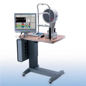
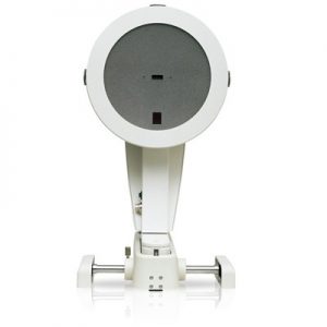
Measurements with Placido Ring Illumination
Thousands of measuring points are used to measure the whole surface of the cornea. A white ring illumination is used for this purpose. An infrared ring illumination is also provided for analysis of the tear film to prevent glare-related reflex secretion.
Measurements with Light Emitting Diodes
The perfect illumination has been integrated for every function of the OCULUS Keratograph® 5M: White diodes for the tear film dynamics, blue diodes for fluo-images, infrared diodes for Meibography.
Technical Data
Features
| Accuracy: |
± 0.1 D |
| Reproducibility |
± 0.1 dpt |
| Number of rings: |
22 |
| Working distance: |
78/100 mm |
| Number of evaluated data points |
22,000 |
| Camera: |
Digital CCD camera |
| Light source: |
Placido illumination: White
Placido illumination: Infrared 880 nm
Fluorescein illumination: Blue 465 nm
Meibography: Infrared 840 nm
Tear film dynamics: White
Pupillometer illumination: Infrared 880 nm |
| Dimensions (W x D x H): |
275 x 320 – 400 mm x 485 – 512 mm |
| Weight |
6.8 kg |
| Minimum PC requirements |
Processor: Intel Core i3 or better, 4GB RAM, Hard drive space: min. 500 GB, Graphic card: Intel HD Graphics 2000 or better, recommended screen resolution: 1920 x 1080 (Full-HD) |

In acc. with the Medical Device Directive 93/42/EEC
 OCULUS is certified in acc. with DIN EN ISO 13485
Software
JENVIS Dry Eye Report NEW
Find the cause of dry eye quickly and reliably. The JENVIS Dry Eye Report is an unique tool for doing this. After measurements are taken using the Keratograph® 5M and the slit lamp, your customer/patient receives an easy-to-grasp print-out. The Dry Eye Report combines screening and consultancy.
TF-Scan Makes the Tear Film Visible
Patient consultation made easy. This software shows the quality and quantity of the tear film.
In cases of dry eye patients and contact lens wearers, the tear film should be examined carefully. Only an intact tear film guarantees contact lens wearing comfort! The Keratograph® 4 measures the tear film breakup time non-invasively (quality assessment). You can show your patient the individual tear film quality using the color maps. In addition, you can take another non-invasive measurement to determine the amount of tear film (tear film quantity).
Tear Film Quality (NIKBUT)
The OCULUS Keratograph® 4 determines the break-up time using the NIKBUT procedure (non-invasive-Keratograph-break-up time).
Meniscus Tear Height Assessment of the Tear Film Quantity
The height of the torn meniscus can be precisely measured with an integrated ruler and various magnification options, and its development along the edge of the bottom lid can be assessed. The results are saved to the patient file.
Lipid Layer Assessment of the Interference Phenomenon
The interference colors of the lipid layer and their structure are made visible and can be recorded. The thickness of the lipid layer is assessed based on the structure and color.
TF Dynamics Assessment of the Particle Flow
The video recording, with up to 32 frames per second, enables the observation of the tear film particle flow, from which conclusions regarding the viscosity of the tear film can be drawn.
Meibo-Scan Meibography of the Top and Bottom Eyelid
The multi-functionality of the new Keratograph® 5M enables even difficult examinations, such as Meibography, to be integrated with ease and efficiency into routine ophthalmological and optometric check-ups. A dysfunction of the Meibom glands is the most common cause of Dry Eye. The associated morphological changes in the glandular tissue can be made visible with the Meibo-Scan.
Pupillometry
Indispensable for:
- Fitting multi-focal contact lenses
- Exact determination of the treatment area for refractive surgery
- Seamless integration into the existing Keratograph® software
- The infrared camera installed in the Keratograph delivers images of the patient’s pupil, which are used as the basis for the measurements
Optional Imaging Software*
The Imaging Software is used to record video and image files. In addition to viewing the videos and single images by themselves, you can also compare the recordings with the simulated fluorescein-images of the measured eye.
OCULUS OxiMap®
The OxiMap® presents a color map of the oxygen transmissibility of soft contact lenses based on the lens power, which is easy to understand – even for your customers!