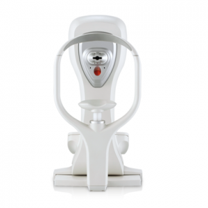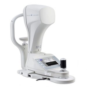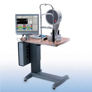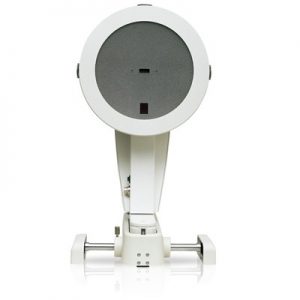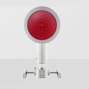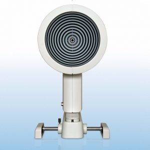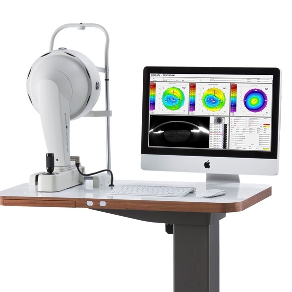


The OCULUS Pentacam® HR is a high-resolution rotating Scheimpflug camera system for anterior segment analysis. It provides crisp images of cornea, iris and crystalline lens. It measures the anterior and posterior corneal topography and elevation, total corneal refractive power, corneal power distribution, automatic chamber angle measurement in 360°, chamber depth and volume, HWTW, corneal and crystalline lens optical opacities. Standard Software: Belin/Ambrosio Enhanced Ecstasia, Holladay Report, Contact Lens Fitting, Cataract package and Refractive Package.
OCULUS Pentacam® HR
The measurement principle
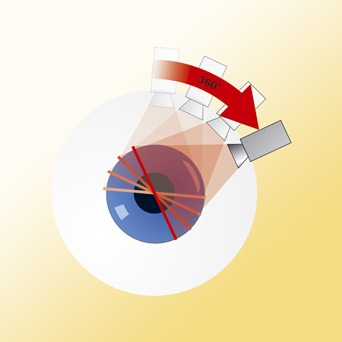 The OCULUS Pentacam is a combined device consisting of a slit illumination system and a Scheimpflug camera which rotates around the eye.
A thin layer within the eye is illuminated through the slit. Being not entirely transparent the cells scatter the slit light. In doing so they create a sectional image which is then photographed in side view by a camera. This camera is oriented according to the Scheimpflug principle, thus creating an image of the illuminated plane which appears completely sharp from the anterior surface of the cornea right up to the posterior surface of the crystalline lens. Swivelling around the eye, the slit-camera device generates a series of radially oriented images of the anterior eye chamber. In the subsequent analysis of the sectional images, tissue boundaries are detected and point clouds are assigned to the various tissue layers (anterior and posterior corneal surfaces, iris, crystalline lens).
The sectional images are saved, corrected in relation to a common reference point and then put together to create a three-dimensional model of the entire anterior eye chamber. This makes it possible to generate reproducible tomographic images of the anterior eye chamber in any desired plane.
After correction for Scheimpflug distortion and light refraction at tissue interfaces the exact of the location of image edge points in the eye is determined by means of ray tracing. Eye movements during image acquisition are captured by a second camera (pupil camera) and also taken into account in the mathematical evaluation. This produces a set of three-dimensional measurement data which gives a precise geometric description of the anterior eye segment. This data in turn can be used to generate data on elevation, curvature, pachymetry, depth of the anterior eye chamber…etc. in the well-known form of colour maps.
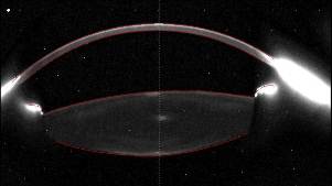 Differences in brightness between individual layers provide an indication of any opacity, since transparent layers result in less slight scattering. This information can be used to assess cataracts.
The OCULUS Pentacam is the only instrument on the market able to perform a precise and complete measurement and analysis of the centre of the cornea. The rotating measurement principle avoids measurement errors that would result from horizontal scanning. Due to the radial orientation of the sectional images the density of data points is greatest at the centre.
This contact-free, hygienic measurement process takes less than two seconds. This is the time it takes to generate 50 sectional images yielding in turn 138,000 true elevation values.
Technical Data
Feature
| Camera | digital CCD camera |
| Light source | blue LEDs (475 nm UV-free) |
| Processor DSP | with 400 mil. operations/s |
| Speed | 100 images in 2 seconds |
| Dimensions | (HxWxD) 535 x 2 80 x 36 0 mm |
| Weight | 9 kg (without elevation table) |
| PC minimum requirements | Pentium IV, 1.5 GHz, Windows XP, 1 GB RAM,
VGA graphic card 1024 x 768 true colour, SB interface |
Measurement Range
| Curvature | 3 – 38 mm (9 – 99 D) |
| Precision | ± 0,1 D |
| Reproducibility | ± 0,1 D |
| Operating distance | 80 mm |
Software
Basic Software:
- Qualitative assessment of the cornea
- Topography and elevation data of the anterior and posterior corneal surface
- Overall pachymetry, absolute and relative
- Glaucoma screening
- Pachymetry based IOP correction
- Chamber angle, -volume and –depth
- Topography based keratoconus detection and classification
- Indices Report to detect abnormalities
- Comparison and differential displays
- Superimposition of Scheimpflug images
- Tomography
- Automatic measurement of the corneal diameter HWTW
Optional software modules
- Software Package Refractive – Software package designed for the Refractive surgeon with the displays and maps listed in the table chart on the previous page.
- Software Package Cataract – Software package designed for the Cataract surgeon with the displays and maps listed in the table chart on the previous page.
- Module Contact lens fitting – Large, unlimited pre-programmed CL database with manual editing function, fluo image simulation, mathematic best fit suggestion.
- Module PNS (Pentacam® Nucleus Staging) and 3D lens density analysis – The density of the crystalline lens is quantified in 3D depending on the selected area. A grading factor called PNS is calculated automatically, independent of any user settings.
- Module Belin/Ambrosio Enhanced Ectasia – This module is based on elevation maps and corneal thickness progression to detect ectasia, designed for virgin eyes.
- Module Holladay Report – This module displays the Equivalent Keratometer Readings that are calculated considering the anterior and posterior curvature of the cornea. The distribution of the EKR’s in different zones is displayed.
- Module phakic IOL simulation and aging prediction – This module simulates iris fixated phakic IOL position in the anterior chamber, predicts aging for up to 50 years after surgery, incl. large pre-programmed database.