Products

/Products/
  • compass

    iCare COMPASS Automated Perimeter with active Retinal Tracking

    Key features

    • Standard automated perimetry
    • Active retinal tracking compensating for poor patient fixation in real-time
    • Auto-focus — no trial lens needed
    • Hygienic design
    • Illustrative fixation analysis; fixation area and plot
    • High-resolution confocal TrueColor imaging of the retina
    • No dilation of pupil needed, the patient can blink freely and the test can be suspended at any time without data loss
    • Ease of use & minimal operator training
  • maia-modello

    iCare MAIA and S-MAIA confocal microperimeters

    Key features
    • 25 Hz eye-tracking with confocal SLO imaging
    • Pre-programmed and custom tests
    • Sensitivity and fixation indexes, illustrative sensitivity, and fixation maps
    • Informative examination and progression reports
    • 2.5 mm minimum pupil size
    • Patients with cataract (up to grade 3+) and media opacities can be examined
    • Auto-focus (from -15D to +10D)
    • Sensitive to functional changes due to macular pathologies or treatments even in early stages (background down to <0.0001 asb, threshold range 36 dB)
    • Simple to use, patients can be tested in less than 3 minutes per eye
    • S-MAIA includes also scotopic testing with cyan and red stimuli
     
  • iCare DRS

    iCare DRS automated digital retinography system

    Key features
    • Fully automated: patient autosensing, auto-alignment, auto-focus, auto flash adjustment, auto-capture
    • Short exam time
    • High quality color and Red-free images
    • Minimal training required
    • Compact and clean design
    • No additional PC required
    • Touch-screen operation
  • The B-Scan Plus’ and B-Scan Plus Vet's proprietary software combined with unique probe electronics make it the only portable High Definition B-Scan. While developing the B-Scan Plus software, Accutome visited many of the top facilities around the world. The results are a reliable and easy-to-use unit, which can quickly scan patients and transport information. Features:
    • 0.015 mm resolution is among the highest in the medical industry.
    • State-of-the-art probe design is sharper, more focused images due to the elimination of signal loss.
    • Contains 4 useful measuring tools along with corresponding A-Vectoring for diagnosing ocular pathologies.
    • The only unit with 2x full image zoom without distortion of real-time or captured scan.
    • Portable probe can plug into any laptop or PC.*
    • Unlimited 34 second film loops.
    • Share Information Easily with adaptable document transfer via EMR, email or printer.
    • Compact file storage is unsurpassed by the competition.
    • Protect your investment with easily upgradeable software.
    • User-friendly interface makes user manuals and quick start guides unnecessary.
    • The B-Scan Plus sofware is now DICOM compatable.
    * Certain Computer Specifications Required.
  • The A-Scan Plus Connect® is designed to meet the demands of today’s refractive cataract surgery.  It is the single most effective solution for measuring and calculating all of your patients and refining your surgical outcomes.  It has the ability to measure most patients and works great with dense cataracts or patients with fixation difficulties. The Connect links directly to a PC, laptop or tablet device making it the ultimate portable solution. Information can easily be uploaded to electronic medical record (EMR) systems. The improved user interface makes entering patient data, capturing the scan and calculating the measurement much faster.* Features:
    • Immersion and Contact Modes.
    • Industry Leading Resolution.
    • User-friendly interface.
    • Less Patient Chair Time – Faster measurement capture*.
    • Automatic Alignment Detection and Sclera Recognition eliminate marginally aligned scans.
    • Portable, Lightweight Design – Plug into any PC, laptop or tablet device.
    • Share information easily – adaptable document transfer via EMR, email or printer.
    • Modern Third and Fourth Generation Formulas including the Hoffer®Q, SRK/T, Holladay, Haigis as well as new post refractive formulas.
    • Optimized Lens Constants for superior surgical outcomes.
    • Unlimited Software Licenses.
    • Streamline your patient data - shares information with B-Scan Plus and UBM Plus.
    *When compared to original A-Scan Plus
  • The intelligence inside

    The Keeler intelligent puff system giving you a quantum leap forwards in accuracy and ease of use. intelliPuff® embodies electronic and optical technology to deliver you the speed, accuracy and ease of use you and your patients deserve.

    Our softest puff ever

    Gentle is understating the new system - and if the patient has high pressures the puff will automatically increase for the next measurement. Kind, intelligent and simple. Technology working for you.

    Desktop or wall mount

    Save valuable space Pulsair intelliPuff® uses less than 50% of the space of conventional non contact tonometers and is the only model that can be wall mounted. Whether you wall mount or prefer to place it on your desk you win the 'space war' every time. No special instrument table is required - put it where you want it. Move it when you want to.  
  • ultra q reflexultra q reflex
    ultra q reflexThe new Ultra Q Reflex™ is the only YAG laser designed to perform both anterior and posterior YAG treatments – and is ideally suited for the treatment of vitreous strands and opacities. 1. Capsulotomy with New Generation IOLs 2. Laser Vitreolysis for the Treatment of Floaters 3. Peripheral Iridotomy for Glaucoma

    Product Information

    1. Optimized Optics for Better Viewing 2. Reflex Technology 3. IOL-Friendly Photodisruption 4. Better Performance Over the Long-Term 5. Pinpoint Precision 6. Accurately Position Optical Breakdown 7. Designed to Maximize Your Workflow 8. Faster Treatment
  • Eyecubedellex Eyecubed

    eye-cubed

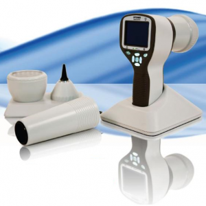
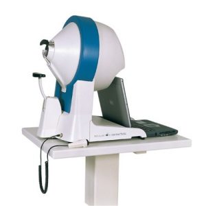
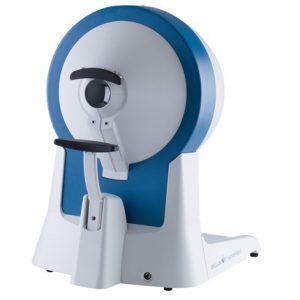
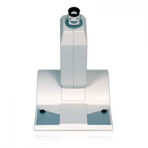
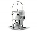
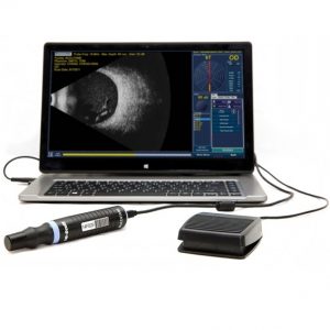
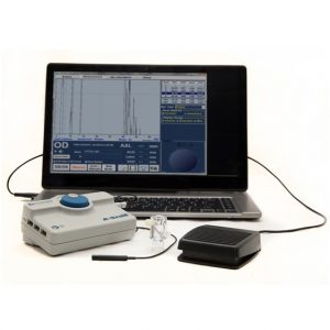
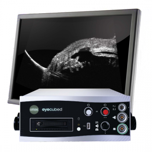
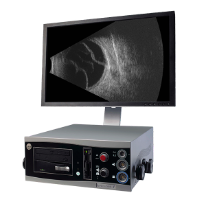
    Eye Cubed™ features real time imaging, advanced movie mode, real-time image enhancement, and a range of self-calibrating "best fit" probes.

    Customized Configuration to Best Meet Your Needs

    With customized configuration of A-Scan and B-Scan modes, Eye Cubed™ covers all of your diagnostic ultrasound needs for both the posterior and anterior segments. Pre-op or post-op, A-Scan or B-Scan, retina or anterior segment: whatever your focus, Eye Cubed™ shows you more, in more detail, than any other device of its kind.

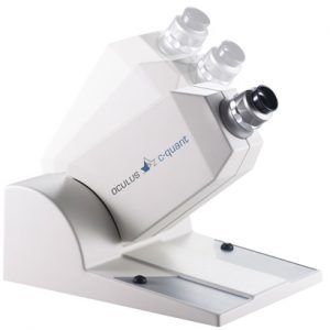
    High Resolution Goes Ultra

    Eye Cubed’s 40 MHz UBM mode allows you to view anterior structures such as the cornea, iris, ciliary body, crystalline and intraocular lens more clearly than ever before. Whether determining the sulcus-to-sulcus measurement for accurate ICL sizing or the angle for potential angle closure and possible YAG laser iridotomy, Eye Cubed’s 40M Hz UBM mode is the gold-standard in high-resolution ultrasound.

    Essential Technology for Your Practice

    Even in an era of high-tech OCT scanning and digital imaging, ultrasound is the only means to obtain a crucial view of the posterior segment when there is a dense cataract or vitreous hemorrhage in the eye – making it one of the most fundamental diagnostic tools in ophthalmology. Detection of disorders like posterior vitreous detachment (PVD) in opaque ocular media is easily achieved with B-Scan ultrasound.

    Innovative Imaging

    Since acquiring Sacramento-based ophthalmic ultrasound pioneers Innovative Imaging Inc. in 2006, Ellex has been working hard to provide ever-increasing value to our Eye Cubed customers – and to develop our ultrasound technology platform. With the support of personalized, clinical ultrasound applications training by expert ecographers, Eye Cubed™ offers a total solution for your practice – and for your patients.
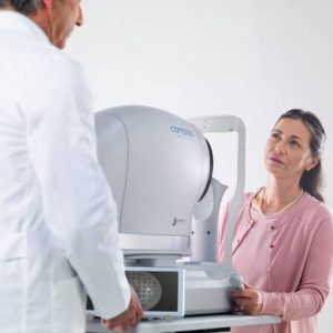
    Ophthalmic Edge

    Visit the comprehensive online diagnostic teaching tool created by Yale Fisher, MD, to access lectures, monographs and cases studies covering a range of diagnostic technologies, including ultrasound and OCT.
  • DIGITAL FUNDUS IMAGING 
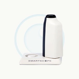
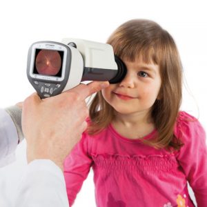
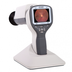
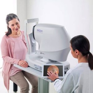
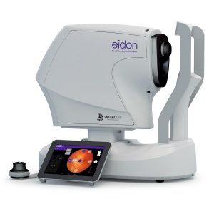
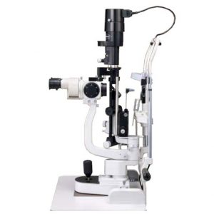
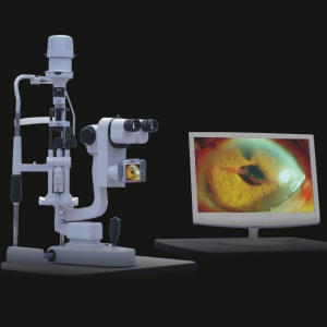
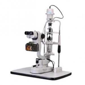
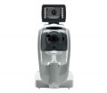
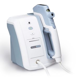
Smartscope PRO camera with Smartscope FA module is a perfect combination of Slit Lamp and hand-held mode providing high quality Fluorescein Angiograms with a wide 40 degrees field of view. 9 internal fixation targets enable peripheral imaging (total FOV 70 * 52 degrees). Smartscope FA is easy to operate and offers fast image capture ability and a detailed view of the entire fluorescein dye circulation dynamics. With a special Slit Lamp Adapter Smartscope FA can be mounted to any Slit Lamp with patient head rest. Wi-Fi enables easy image transfer to PC, laptop, tablet or mobile devices. With Optomed Workstation software (sold separately) the images can be viewed, shared and stored locally and sent directly to a DICOM compatible system (PACS or other) of the hospital network.
  • optomed smartscope prooptomed smartscope pro
    DIGITAL FUNDUS IMAGING Optomed Smartscope® PRO is the next generation hand-held medical camera that provides high image quality which fulfills international ISO 10940 fundus camera standard requirements. Interchangeable modular optics make the camera even more versatile. The lightweight camera incorporates advanced technology in a portable design. Smartscope PRO camera provides accurate and silent autofocus which makes image capturing quick and easy. The camera is also equipped with Wi-Fi for advanced image transfer to any PC or mobile device. The Smartscope PRO comes with Patient Data Management software (sold separately) with full DICOM and PACS compliance to ensure seamless interoperability with hospital networks and practice management systems.
  • Our cutting edge retinal imaging technology is customized to fit the needs of your practice. The next generation of Optos technology, Daytona has been scaled to accommodate smaller office spaces while providing ultra-high resolution imaging, and adding ultra-widefield autofluorescence capabilities. Features:
    • Non-mydriatic ultra-high resolution images in under a second, through 2 mm pupils and many cataracts
    • Red and green lasers; each wavelength provides information for interpretation and diagnosis. Channels can be viewed separately:
      • Green (532 nm) “red-free” visualizes the sensory retina to the RPE
      • Red (635 nm) shows deeper structures of the retina (RPE to Choroid)
    • Ultra-widefield autofluorescence imaging with green laser light displays lipofuscin in the RPE
    • Images are available immediately and stored electronically for future comparison or telehealth applications
    • Innovative software tools enhance image evaluation
    • DICOM compatible
  • Optos-CaliforniaOptos-California
    With California, Optos has incorporated  new hardware and software technology enabling practitioners  to see more, discover more and effectively treat more ocular pathology thus promoting patient health.  We are committed to further strengthening our clinical evidence while demonstrating the importance of imaging the entire retina. We have created three versions of California to be customizable for use in multiple eyecare settings. California includes a new UWF optomap imaging modality; Indocyanine Green angiography (icg) while retaining:
    • Composite color
    • Red-free
    • Autofluorescence (af)
    • Fluorescein angiography (fa)
    Images are now presented in ProView which displays optomap in a consistent geometry that accurately represents anatomical features in the retina. Further, ProView enables automatic image registration for disease tracking over time, and inter-modality image comparison. New proprietary optical hardware optimizes and maintains resolution of the optomapimages throughout the scan of the retina resulting in more clarity in the far periphery. Image overlay enables comparison between composite color images and red-free, affa, or icg images.  Additionally, comparisons can be made between different images or different dates by scrolling through all stored images.
  • Tango_SLTtango_ellex
    tangoTango™ combines a full-featured SLT laser with a powerful, precise YAG laser, helping you to better manage both OAG and ACG patients, while also providing a highly effective solution for capsulotomy procedures. Engineered and built by Ellex, Tango™ brings together 30 years of manufacturing expertise and cutting-edge technology to deliver consistently better treatment results. Featuring a proprietary, patented dual-mode laser cavity, which allows you to instantly – and reliably – switch between SLT and YAG modes, combined with a custom-designed power supply that maintains optimum system stability for greater shot-to-shot consistency, Tango™ allows you to perform SLT and YAG treatments with improved accuracy and greater efficacy over the long-term.  

    Product Information

    1.  Optimized for Anterior Segment Viewing
    2. IOL-Friendly Photodisruption
    3. Better Performance Over the Long-Term
    4. Faster Treatment
    5. Pinpoint Precision
    6. Accurately Position Optical Breakdown
    7. Designed to Maximize Your Workflow
    8. Greater Efficiency at the Touch of a Button
    9. A Wider View
    10. Configured to Meet Your Needs
  • optomed_M5_Standard
    DIGITAL FUNDUS IMAGING Portable Non-mydriatic eye fundus examination Optomed Smartscope M5 is a digital medical camera that provides general, ophthalmoscopic, otoscopic and dermatoscopic imaging with one hand-held device. This multipurpose digital imaging device weights only 400g and powered with a battery it gives you the freedom to move around and take the device with you to any location. Digital still and video images created with Optomed Smartscope M5 allow making accurate first diagnosis and planning consistent follow-up treatment. Optomed Smartscope M5 is easily adopted into daily examination routines and connectivity to any patient database system enables fluent image data sharing e.g. for consultation purposes. The device comes with a 2GB SD memory card and pictures can be easily transferred to your PC by placing the device to its cradle which connects to the PC via USB connection. The 5 megapixel sensor and up to 2560 x 1920 pixels resolution guarantee the quality of your pictures. Each of our 4 attachable optics modules has its own light source and adjustable illumination levels. Optics are fast and easy to attach and detach with bayonet connectors. The advances in M5 compared to M3-2 are new sensor enabling improved picture quality, higher battery capacity and keyboard lights that make using the device in a dark room easier. Optomed Smartscope M5 offers non-mydriatic fundus imaging without the need for dilation drops with EY2 and EY3 optics modules.
  • iCare EIDON widefield TrueColor Confocal fundus imaging system

    Key features
    • Multiple imaging modalities including TrueColor, blue, red and Red-Free and infrared confocal images
    • Widefield, ultra-high-resolution imaging
    • Capability to image through cataract and media opacities
    • Dilation-free operation (minimum pupil 2.5 mm)
    • Flexibility of fully automated and fully manual mode
    • All-in-one compact design, no additional PC required
  • Fly_Stereopsis_Test_LEA_lg

    Designed to rapidly test for amblyopia and strabismus using gross thru fine stereopsis (4800 to 20 seconds of arc). Graded circle test from 400 seconds now down to 20 seconds with no monocular clues. New improved booklet has answer key on back cover and includes polarized viewers.

    • Graded circle test from 400 seconds now down to 20 seconds
    • No monocular clues with NEW technology
    • New improved booklet
    • Answer key on back cover
    • Includes both adult and pediatric polarized viewers
  • Random_Dot_Stereopsis_Test_lg
    Designed to rapidly test for amblyopia and strabismus in early and non-readers and non-verbal children & adults. Expanded Random Dot LEA Symbols Test (500, 250, 125, 63 seconds of arc). Graded circle test now down to 12.5 seconds with no monocular clues. New improved booklet has answer key on back cover and includes polarized viewers.
    • Expanded Random Dot Lea Symbols Test
    • Graded circle test now down to 12.5 seconds
    • No monocular clues with NEW technology
    • New improved booklet
    • Answer key on back cover
    • Includes both adult and pediatric polarized viewers
  • Random_Dot2_lg
    Designed to rapidly test for amblyopia and strabismus in early and non-readers and non-verbal children& adults. Expanded Random Dot Lea Symbols Test (500, 250, 125 seconds of arc). Graded circle test now down to 12.5 seconds with no monocular clues. New improved booklet has answer key on back cover and includes polarized viewers.
    • Expanded Random Dot Lea Symbols Test
    • Graded circle test now down to 12.5 seconds
    • No monocular clues with NEW technology
    • New improved booklet
    • Imprinted images inside booklet for matching
    • Answer Key on back cover
    • Includes both adult and pediatric polarized viewers
  • 1015VAC_lg
    Designed to rapidly test for amblyopia and strabismus in early and non-readers and non-verbal children and adults. Choice of dot size acuity (standard and low vision). Includes adult and new child-friendly pediatric polarized viewers.
    • Choice of dot size acuity
    • New improved test frames
    • Includes both adult and pediatric polarized viewers
  • Butterfly_Stereopsis_Test_lg
    Designed to rapidly test for amblyopia and strabismus using gross thru fine stereopsis (2500 to 20 seconds of arc). Graded circle test from 400 seconds now down to 20 seconds with no monocular clues. New improved booklet has answer key on back cover and includes polarized viewers.
    • Graded circle test from 400 seconds now down to 20 seconds
    • No monocular clues with NEW Technology
    • New improved booklet
    • Answer key on back cover
    • Includes both adult and pediatric polarized viewers
  • ri-scope Ophthalmoscoperi-scope Ophthalmoscope
    All ri-scope® L ophthalmoscopes with modular design impress with their high-performance optics with aspherical condenser lens with reduced-refl ection view even with small pupils and through improved ergonomics. The new ri-scope® L ophthalmoscopes have a thumbrest for securing the ophthalmoscope, which enables easy control of all the elements with one finger. ri-scope® L2 The enhanced basic model with separately engageable red-free, blue  and polarization filters for each aperture.
    • Diopter wheel with 29 corrective lenses
    • Plus 1-10, 12, 15, 20, 40; Minus 1-10, 15, 20, 25, 30, 35
    • Easy-to-operate aperture hand-wheel with semi-circle, small/medium/large circle, fixation star, and slit
    • Includes filter wheel engageable for all apertures with symbol display, red-free filter, blue filter and polarization filter
    • Optimized high-performance optics with aspherical condenser lens
    • Parallel beam path
    • Dust-proof
  • ri-scope _retinoscoperi-scope retinoskop
    The ri-scope® retinoscope (skiascope) measures the refractive power of the eye.  Errors of refraction, such as short and long-sightedness, as well as astigmatism can be recognised and determined. Choice of XL 3.5 V xenon lamp or HL 2.5 V halogen lamp.
  • Inami Slit Lamp
    The L-0189 5 STEPS MAGNIFICATION SLIT LAMP provides practitioners with the utmost satisfaction thanks to its excellent image clearness and fine design.
  • iCare EIDON AF blue autofluorescence confocal fundus imaging system

    Key features

    • TrueColor, blue autofluorescence, infrared and RGB channels confocal images
    • Panoramic view of the retinal autofluorescence (up to 110°)
    • High details and contrast in the same device
    • Short exam time and enhanced patient comfort
    • Easy to use, speeds up patient workflow
  • SLM-4 ER Digital Slit Lamp
    Features Binocular linkage Galileo style Five Steps Rotation magnification Large depth of field and high definition optical system Digital camera: image captured by button on handle Image system software based on Windows 2000/XP (optional)
  • SLM-3 ER Digital Slit Lamp
    Professional image analyzing, processing and management system Professional optics with high definition digital SLR camera (16.2 Mega Pixels) Special case storage system Professional software with high definition video function Background illumination system
  • SLM-3 ER-E Digital Slit Lamp
    SLR camera (24.1 Mega Pixels); Special case storage system Professional image analyzing, processing and management system; Flash illumination compensation system High definition video function Background illumination system Power box supplies camera power directly. without charging battery Super optical Top definition eyepiece Adjusting the illumination by Joystick, it is much easier to use. Economic, high quality, low maintenance and depreciation New function: Illumination control by Joystick Eyepiece correction (Same focus) Automatic image optimization (such as color, brightness) Filter for corneal staining (Five different color is more easier for doctor to get best images)
  • SLM-3 ER (CCD) Digital Slit Lamp
    Professional image analyzing, processing and management system Professional optics with CCD Special case storage system Professional software with video function Background illumination system Flash function
  • SLM-2 ER-E Slit Lamp
    Galileo parallel design for using, five magnifications 14mm aperture provides broader and clearer field of view High sensitivity professional objective lens German industrial bulb, high brightness without harsh, quick heat dispersion, also provide the low heating, high brightness and even lighting. International standard accessory interface which can connect kinds of ocular posterior segment laser adapter and foldman tonometer Digital upgrading interface. Can upgrade to digital slit lamp Excellent optics performance, versatility and ease of use Stereo variator, besretinal view even through small pupils Ergonomic, wide aperture optics for fatigue free daily use
  • simple bc trial frameBicoh_BC Trial Frame
    FEATURES: Fixed P.D. PD Size: 13 sizes from 48mm to 72mm, with 2mm interval. Light weight Only 36g without lenses Axis of astigmatism Provides each 5° Nose pad Adjustable in height Temple With soft temple tip. Available in NON SLIP temple tips (Optional) Color Black (PD 48-52 type are only blue or pink)    
  • bicoh universal trial frame 674Bicoh_trial farme MD674
    FEATURES: Dual Hold Temple Tip (Side & downward direction) Half Rim Type (For Progressive Lens available) Fixed P.D. PD Size: 11 sizes from 54mm to 74mm, with 2mm interval. Light weight Less Than 50g without lenses Axis of astigmatism Provides each 5° Nose pad Adjustable in width and height Temple Adjustable in angle and length, with NON SLIP temple tips.  
  • teaser_lupenbrilleoculus_lupenbrille
    For decades, OCULUS binocular loupes have been a tried and tested system worldwide for precision work in many fields, such as medicine, art, research, technology and many others. These magnifying loupes are ideal for low vision patients to improve the quality of life. They make stereoscopic near vision possible and provide a high rate of mobility.
    Thanks to the continuous further development of these powerful binocular loupes, a complete system offering the following features is now available:
    • The high-quality OCULUS optic ensures bright images, true-color and depth of field.
    • The large diameter exit pupil makes binocular adjustment easy for optimal vision.
    • The low weight and the ergonomically designed system carriers prevent strain, even for longer periods of wear.
    • The ability to adjust the pupil distance and change the angle of convergence allows optimal adaptation to the wearer’s individual needs.
    • For room orientation purposes, the magnifying lenses can be flipped up, without having to change the system settings.
    • A clear view through the metal frame is maintained.
    • Corrective or protective lenses can be inserted into the metal frame.
    • Optimal near vision adapters enable wearers to use standard multi-focal lenses. The modular concept of the 1.8x binocular loupe allows optics with other magnifications to be mounted quickly and easily, for flexible adjustment to changing requirements.
  • oculus_binoptometer4p_mit_tabletteaser_binoptometer_4p_produkt_02
    The OCULUS Binoptometer® 4P is a professional screening instrument for testing all relevant visual functions. The used future-oriented technology allows an almost unlimited representation of various vision tests. Vision-screening instrument with contrast sensitivity testing and optional mesopic vision and glare sensitivity testing.

    New Test Presentation

    Vision tests are presented on a high-resolution micro-color display, allowing a virtually unlimited presentation of vision tests.

    Unbeatable

    Contrast sensitivity, optional mesopic vision and glare sensitivity testing with just one instrument.

    Ergonomic

    Convenient height adjustment range of 4.13 inch (10.5 cm). In addition, the viewing angle is fully adjustable up to a 35° downgaze, thus enabling testing to be done ergonomically with any eyeglasses.
  • oculus_centerfield_moderne_verbindungoculus_centerfield_ergonomisches_design
    Thanks to its ergonomic design, user-friendly program navigation and ease of operation, the OCULUS Centerfield® 2 Perimeter has proven itself to be an invaluable instrument in the Occupational Health area. The unit performs static perimetry up to 70° eccentricity.  It also meets the requirements of the German Ophthalmological Society’s (DOG) Road Traffic Commission for conducting visual field testing in accordance with the regulations for the issuance of driver’s licenses and the Guidelines G25 and G41 issued by the German Institute for Occupational Health and Safety.

    Ergonomic Design

    The self-contained design and light-shielded viewer allow examinations to be conducted in normally lit rooms.  Its robustness and light weight make the OCULUS Centerfield® 2 Perimeter the ideal unit for portable use, which is often a necessity in the Occupational Health area.

    Reliable Results

    The results of the perimetric measurements are summarized in a clearly structured printout. Areas of abnormality can be quickly recognized and re-examination of these areas independent of the test point grid gives the diagnostic analysis even greater reliability.

    Modern Connectivity

    The OCULUS Centerfield® 2 Perimeter can be operated via the USB port of a notebook or PC. Together with the supplied device software, this modern solution allows you to fully utilize all of the benefits of today’s IT systems, especially the network connectivity. This guarantees both safe storage of examination data and quick access to that data when needed.
  • oculus - corvisoculus_corvis_tonometer
    The revolutionary OCULUS Corvis® STL records the reaction of the cornea to a defined air pulse with a newly developed high-speed Scheimpflug-camera that takes over 4,300 images per second. IOP and corneal thickness can be measured with great precision on the basis of the Scheimpflug images.

    Corneal Visualization Scheimpflug Technology

    The revolutionary OCULUS Corvis® STL records the reaction of the cornea to a defined air pulse with a newly developed high-speed Scheimpflug-camera that takes over 4,300 images per second. IOP and corneal thickness can be measured with great precision on the basis of the Scheimpflug images.

    Measurement and Display Options

    • IOP measurement
    • Measurement of the corneal thickness
    • Scheimpflug images of the 1st and 2nd applanation of the cornea
    • Slow-motion video of the corneal deformation as a result of the air pulse
    • Non-contact tonometer in combination with an ultra-high-speed camera for visualization of the deformation of the cornea in reaction to an air pulse (4,330 frames / sec.)
  • oculus_cquantoculus_cquant_1
    With this product development, OCULUS is once again living up to its motto, "We focus on progress”. The OCULUS C-Quant makes it possible to measure the amount of straylight in a human eye individually and reliably. The effect of increased straylight in the human eye can be compared to looking through a dirty windshield.