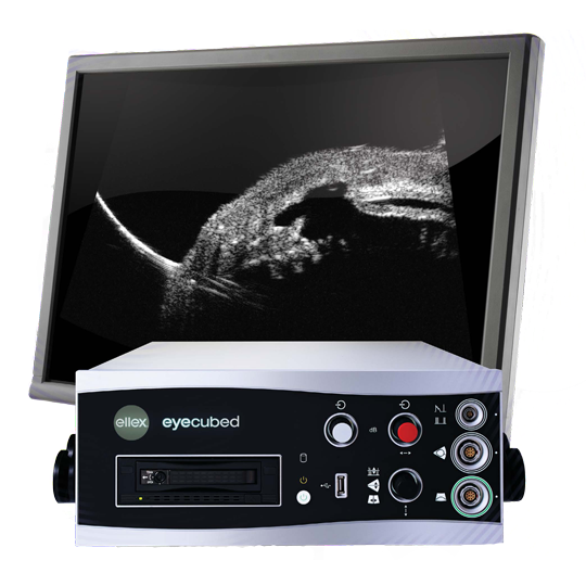
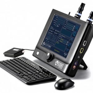
1. Customized Configuration
Customized configuration of A-Scan and B-Scan modes ensures that Eye Cubed™ meets all of your needs for both the posterior and anterior segments.
- 40 MHz UBM B-Scan mode delivers accurate measurement and evaluation of the iris, angle and cilliary body, including sulcus-to-sulcus (ICL sizing) and IOL haptic placement.
- 10 MHz Posterior B-Scan mode produces the subtlest vitreous echoes, offering unparalleled distinction between the retina, choroid and sclera, as well as the vitreo retinal junction.
- Biometry A-Scan delivers ultra-precise axial length measurement with faster, easier image acquisition in real-time movie mode.
- Standardized Diagnostic A-Scan enables precision tissue differentiation.
2. Highest Signal-to-Noise Ratio
The unique amplifier and probe design of Eye Cubed™ provides for the industry’s highest signal-to-noise ratio. That is, Eye Cubed™ delivers substantially more ultrasonic data per second than any other system. Because noise is reduced to a minimum, details of even the finest ocular structures become visible – including blood and inflammatory cells.
3. Advanced Movie Technology
Eye Cubed’s advanced movie technology greatly improves the diagnostic capability and speed of each exam, allowing you to capture up to 20-second movies. Review these movies frame-by-frame in order to reveal greater detail, or play them back in full movie mode.
4. High Speed Imaging for Real-Time Display
With an image acquisition rate of up to 25 frames-per second, Eye Cubed™ provides the fastest image-sampling rate available. This speed creates a real-time view of detailed ocular activity, including blood cell movement and membrane behavior.
5. Intuitive User-Friendly Software
Eye Cubed™ incorporates a number of features designed to accelerate practice workflow, including improved export and import functionality and expanded measurement options. Intuitive and easy-to-use, the software incorporates a multilingual user interface, full-screen scan viewing and customized report capability.
6. Custom Velocity Settings for Greater Precision
In addition to pre-programmed velocities for phakic, aphakic and 4 types of pseudo-phakic eyes, Eye Cubed™ enables you to adjust for all particular cases and program velocities accordingly.
7. DICOM Connectivity
DICOM connectivity (optional license) streamlines the examination process, from start to finish:
- Load patient work list from the EMR system and select patient record
- Perform examination
- Store examination reports to the network (EMR system or Picture Archiving and Communication System – PACS)
8. Real-Time Image Processing
A series of four image processing algorithms allow you to enhance each scan in real-time – not after the fact. These algorithms are optimized for use in ophthalmic echography and have been developed by Ellex’s in-house software engineering team to deliver greater image control. Choose your preferred interpolation method from a number of options, including:
- Lowest neighbour
- Linear interpolation
- Bilinear interpolation
- Cubic interpolation
9. Sensitive Scan™ Transmit Energy Control for Greater Image Detail
Featuring Sensitive Scan™ technology, Eye Cubed™ enables you to adjust the probe transmit energy to ensure optimal tissue sensitivity. This gives you the ability to discern between the finest ocular structure, such as subtle vitreous opacities and sub retinal fluids.
Specifications
| Network and Connectivity |
Electrical Requirements |
| Six USB 2.0 ports for memory sticks and peripherals |
Power supply: 100-240 VAC auto-ranging |
| Fully network and printer-ready (gigabit Ethernet) |
Frequency: 50/60 Hz |
| Purpose-built Windows embedded operating system |
Input power: 220 VA |
| Multilingual user interface |
System size: 15.5 x 17 x 6.5 inches (39 x 43 x 16.5 cm) |
|
Weight: 26 lbs. (12kg) |
| Data Management |
Hardware Features |
| DICOM Connectivity
– Verification of multiple concurrent DICOM connections to other Application Entities (AEs)
– Query / retrieval of modality work list (patient data from Electronic Medical Records – EMR)
– Storage of DICOM objects to EMR / Picture Archiving and Communication Systems (PACS)
|
Footswitch control (scan start, scan stop, etc.) |
| Customized report capability |
Removable one-terabyte hard drive |
| Data archiving and image / movie export capability |
Wide screen, 1920 x 1200 high-resolution monitor |
| B-Scan Modes |
A-Scan Modes |
| Four sets of electronic distance measurement calipers with variable velocity |
IOL power calculations and analysis:
– Holladay-I
– SRK-T
– Haigis (optional)
– Hoffer-Q |
| Two sets of electronic angle measurement callipers (variable velocity) |
Movie sequence adjustable up to 5 seconds |
| Text annotations |
50 frames-per-second image acquisition rate |
| Movie sequence capture up to 20 seconds– Real-time image viewing and editing capability |
|
10 MHz Posterior Segment
|
Axial Length Biometry A-Scan
|
| 25 frames-per-second image acquisition rate |
Immersion or contact method |
| 10-second movie loop capability |
Solid focused probe with internal fixation light |
| Sealed probe |
Probe frequency: 10 MHz |
| Adjustable transmit gain (27-90 dB) |
Image depth: 40 mm |
| Adjustable dynamic range (Log, S1, S2, S3) |
Points on x-axis: 2048 |
| Axial resolution: 50 microns |
8 bit resolution |
| Lateral resolution: 100 microns |
Steps of resolution: 256 |
| Scanning angle: 52 degrees |
Measurement accuracy: 50 microns inherent, 100 microns clinical |
| Image depth (displayed image): 48 mm |
Automatic or manual scan acquisition |
|
Built-in pattern recognition with automatic scleral echo detection |
40 MHz UBM Wide-Field Anterior Segment
|
Standardized Diagnostic A-Scan
|
| 13 frames-per-second image acquisition rate |
Measurement accuracy: 50 microns inherent, 100 microns clinical |
| 20-second movie loop capability |
Two caliper measurements displayed in mm with variable velocities |
| Adjustable transmit gain (minimum to 0 dB) |
Tissue sensitivity value stored in memory with reset function |
| Adjustable dynamic range (Log, S1, S2, S3) |
Probe frequency: 8 MHz parallel beam |
| Axial resolution: 23 microns |
|
| Lateral resolution: 33 microns |
|
| Scanning angle: 30 degrees |
|
| Image depth (displayed image): 11.9 mm |
|
| Image width at focal zone: 15-18 mm |
|
| Focal range: 10.5 – 14.5 mm |
|
Specifications are subject to change without notice.
Image Gallery
1. Melanoma – Choroid
10MHz Posterior Segment B-Scan: Extraocular local recurrence adjacent to the optic nerve following plaque radiotherapy of a medial choroidal melanoma in the right eye.
Images courtesy of Bertil Damato, PhD, FRCOphth, Royal Liverpool University Hospital, United Kingdom.
-
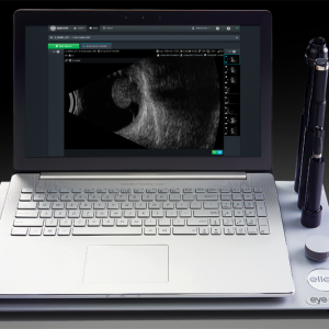
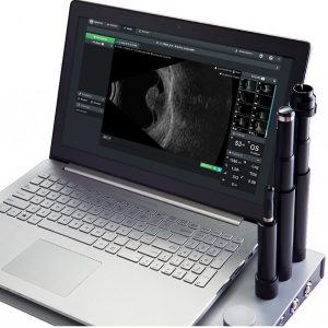
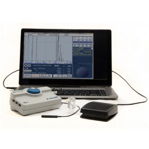
Eye Cubed™ features real time imaging, advanced movie mode, real-time image enhancement, and a range of self-calibrating "best fit" probes.
Customized Configuration to Best Meet Your Needs
With customized configuration of A-Scan and B-Scan modes, Eye Cubed™ covers all of your diagnostic ultrasound needs for both the posterior and anterior segments. Pre-op or post-op, A-Scan or B-Scan, retina or anterior segment: whatever your focus, Eye Cubed™ shows you more, in more detail, than any other device of its kind.
High Resolution Goes Ultra
Eye Cubed’s 40 MHz UBM mode allows you to view anterior structures such as the cornea, iris, ciliary body, crystalline and intraocular lens more clearly than ever before. Whether determining the sulcus-to-sulcus measurement for accurate ICL sizing or the angle for potential angle closure and possible YAG laser iridotomy, Eye Cubed’s 40M Hz UBM mode is the gold-standard in high-resolution ultrasound.
Essential Technology for Your Practice
Even in an era of high-tech OCT scanning and digital imaging, ultrasound is the only means to obtain a crucial view of the posterior segment when there is a dense cataract or vitreous hemorrhage in the eye – making it one of the most fundamental diagnostic tools in ophthalmology. Detection of disorders like posterior vitreous detachment (PVD) in opaque ocular media is easily achieved with B-Scan ultrasound.
Innovative Imaging
Since acquiring Sacramento-based ophthalmic ultrasound pioneers Innovative Imaging Inc. in 2006, Ellex has been working hard to provide ever-increasing value to our Eye Cubed customers – and to develop our ultrasound technology platform. With the support of personalized, clinical ultrasound applications training by expert ecographers, Eye Cubed™ offers a total solution for your practice – and for your patients.
Ophthalmic Edge
Visit the comprehensive online diagnostic teaching tool created by Yale Fisher, MD, to access lectures, monographs and cases studies covering a range of diagnostic technologies, including ultrasound and OCT." href='https://www.ophthalmic.com.sg/wp-content/uploads/2016/09/01-Melanoma-Choroid-002-lg-1024x686-e1472908393124.jpg'>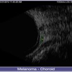
-
Eye Cubed™ features real time imaging, advanced movie mode, real-time image enhancement, and a range of self-calibrating "best fit" probes.
Customized Configuration to Best Meet Your Needs
With customized configuration of A-Scan and B-Scan modes, Eye Cubed™ covers all of your diagnostic ultrasound needs for both the posterior and anterior segments. Pre-op or post-op, A-Scan or B-Scan, retina or anterior segment: whatever your focus, Eye Cubed™ shows you more, in more detail, than any other device of its kind.
High Resolution Goes Ultra
Eye Cubed’s 40 MHz UBM mode allows you to view anterior structures such as the cornea, iris, ciliary body, crystalline and intraocular lens more clearly than ever before. Whether determining the sulcus-to-sulcus measurement for accurate ICL sizing or the angle for potential angle closure and possible YAG laser iridotomy, Eye Cubed’s 40M Hz UBM mode is the gold-standard in high-resolution ultrasound.
Essential Technology for Your Practice
Even in an era of high-tech OCT scanning and digital imaging, ultrasound is the only means to obtain a crucial view of the posterior segment when there is a dense cataract or vitreous hemorrhage in the eye – making it one of the most fundamental diagnostic tools in ophthalmology. Detection of disorders like posterior vitreous detachment (PVD) in opaque ocular media is easily achieved with B-Scan ultrasound.
Innovative Imaging
Since acquiring Sacramento-based ophthalmic ultrasound pioneers Innovative Imaging Inc. in 2006, Ellex has been working hard to provide ever-increasing value to our Eye Cubed customers – and to develop our ultrasound technology platform. With the support of personalized, clinical ultrasound applications training by expert ecographers, Eye Cubed™ offers a total solution for your practice – and for your patients.
Ophthalmic Edge
Visit the comprehensive online diagnostic teaching tool created by Yale Fisher, MD, to access lectures, monographs and cases studies covering a range of diagnostic technologies, including ultrasound and OCT." href='https://www.ophthalmic.com.sg/wp-content/uploads/2016/09/01-Melanoma-Choroid-001-lg-1024x686-e1472908407379.jpg'>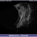
2. Melanoma – Inferonasal
10MHz Posterior Segment B-Scan: Inferonasal melanoma in the left eye measuring 15.7 by 9.5 by 5.2 mm extending to pars plana.
Images courtesy of Bertil Damato, PhD, FRCOphth, Royal Liverpool University Hospital, United Kingdom.
-
Eye Cubed™ features real time imaging, advanced movie mode, real-time image enhancement, and a range of self-calibrating "best fit" probes.
Customized Configuration to Best Meet Your Needs
With customized configuration of A-Scan and B-Scan modes, Eye Cubed™ covers all of your diagnostic ultrasound needs for both the posterior and anterior segments. Pre-op or post-op, A-Scan or B-Scan, retina or anterior segment: whatever your focus, Eye Cubed™ shows you more, in more detail, than any other device of its kind.
High Resolution Goes Ultra
Eye Cubed’s 40 MHz UBM mode allows you to view anterior structures such as the cornea, iris, ciliary body, crystalline and intraocular lens more clearly than ever before. Whether determining the sulcus-to-sulcus measurement for accurate ICL sizing or the angle for potential angle closure and possible YAG laser iridotomy, Eye Cubed’s 40M Hz UBM mode is the gold-standard in high-resolution ultrasound.
Essential Technology for Your Practice
Even in an era of high-tech OCT scanning and digital imaging, ultrasound is the only means to obtain a crucial view of the posterior segment when there is a dense cataract or vitreous hemorrhage in the eye – making it one of the most fundamental diagnostic tools in ophthalmology. Detection of disorders like posterior vitreous detachment (PVD) in opaque ocular media is easily achieved with B-Scan ultrasound.
Innovative Imaging
Since acquiring Sacramento-based ophthalmic ultrasound pioneers Innovative Imaging Inc. in 2006, Ellex has been working hard to provide ever-increasing value to our Eye Cubed customers – and to develop our ultrasound technology platform. With the support of personalized, clinical ultrasound applications training by expert ecographers, Eye Cubed™ offers a total solution for your practice – and for your patients.
Ophthalmic Edge
Visit the comprehensive online diagnostic teaching tool created by Yale Fisher, MD, to access lectures, monographs and cases studies covering a range of diagnostic technologies, including ultrasound and OCT." href='https://www.ophthalmic.com.sg/wp-content/uploads/2016/09/02-Melanoma-Inferonasal-001-lg-1024x686-e1472908428633.jpg'>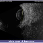
-
Eye Cubed™ features real time imaging, advanced movie mode, real-time image enhancement, and a range of self-calibrating "best fit" probes.
Customized Configuration to Best Meet Your Needs
With customized configuration of A-Scan and B-Scan modes, Eye Cubed™ covers all of your diagnostic ultrasound needs for both the posterior and anterior segments. Pre-op or post-op, A-Scan or B-Scan, retina or anterior segment: whatever your focus, Eye Cubed™ shows you more, in more detail, than any other device of its kind.
High Resolution Goes Ultra
Eye Cubed’s 40 MHz UBM mode allows you to view anterior structures such as the cornea, iris, ciliary body, crystalline and intraocular lens more clearly than ever before. Whether determining the sulcus-to-sulcus measurement for accurate ICL sizing or the angle for potential angle closure and possible YAG laser iridotomy, Eye Cubed’s 40M Hz UBM mode is the gold-standard in high-resolution ultrasound.
Essential Technology for Your Practice
Even in an era of high-tech OCT scanning and digital imaging, ultrasound is the only means to obtain a crucial view of the posterior segment when there is a dense cataract or vitreous hemorrhage in the eye – making it one of the most fundamental diagnostic tools in ophthalmology. Detection of disorders like posterior vitreous detachment (PVD) in opaque ocular media is easily achieved with B-Scan ultrasound.
Innovative Imaging
Since acquiring Sacramento-based ophthalmic ultrasound pioneers Innovative Imaging Inc. in 2006, Ellex has been working hard to provide ever-increasing value to our Eye Cubed customers – and to develop our ultrasound technology platform. With the support of personalized, clinical ultrasound applications training by expert ecographers, Eye Cubed™ offers a total solution for your practice – and for your patients.
Ophthalmic Edge
Visit the comprehensive online diagnostic teaching tool created by Yale Fisher, MD, to access lectures, monographs and cases studies covering a range of diagnostic technologies, including ultrasound and OCT." href='https://www.ophthalmic.com.sg/wp-content/uploads/2016/09/02-Melanoma-Inferonasal-002-lg-1024x686-e1472908439767.jpg'>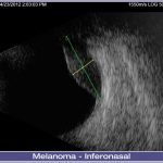
-
Eye Cubed™ features real time imaging, advanced movie mode, real-time image enhancement, and a range of self-calibrating "best fit" probes.
Customized Configuration to Best Meet Your Needs
With customized configuration of A-Scan and B-Scan modes, Eye Cubed™ covers all of your diagnostic ultrasound needs for both the posterior and anterior segments. Pre-op or post-op, A-Scan or B-Scan, retina or anterior segment: whatever your focus, Eye Cubed™ shows you more, in more detail, than any other device of its kind.
High Resolution Goes Ultra
Eye Cubed’s 40 MHz UBM mode allows you to view anterior structures such as the cornea, iris, ciliary body, crystalline and intraocular lens more clearly than ever before. Whether determining the sulcus-to-sulcus measurement for accurate ICL sizing or the angle for potential angle closure and possible YAG laser iridotomy, Eye Cubed’s 40M Hz UBM mode is the gold-standard in high-resolution ultrasound.
Essential Technology for Your Practice
Even in an era of high-tech OCT scanning and digital imaging, ultrasound is the only means to obtain a crucial view of the posterior segment when there is a dense cataract or vitreous hemorrhage in the eye – making it one of the most fundamental diagnostic tools in ophthalmology. Detection of disorders like posterior vitreous detachment (PVD) in opaque ocular media is easily achieved with B-Scan ultrasound.
Innovative Imaging
Since acquiring Sacramento-based ophthalmic ultrasound pioneers Innovative Imaging Inc. in 2006, Ellex has been working hard to provide ever-increasing value to our Eye Cubed customers – and to develop our ultrasound technology platform. With the support of personalized, clinical ultrasound applications training by expert ecographers, Eye Cubed™ offers a total solution for your practice – and for your patients.
Ophthalmic Edge
Visit the comprehensive online diagnostic teaching tool created by Yale Fisher, MD, to access lectures, monographs and cases studies covering a range of diagnostic technologies, including ultrasound and OCT." href='https://www.ophthalmic.com.sg/wp-content/uploads/2016/09/02-Melanoma-Inferonasal-003-lg-1024x686-e1472908451195.jpg'>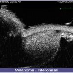
3. Melanoma – Inferotemporal Ciliary Body
10MHz Posterior Segment B-Scan: A left inferotemporal ciliary body melanoma measuring 13.5 x 11.4 x 5.5mm. (Note: Ciliary body melanomas can have cystic spaces or pseudocystic spaces if there are areas of necrosis or amelanotic melanoma.)
Image courtesy of Bertil Damato, PhD, FRCOphth, Royal Liverpool University Hospital, United Kingdom.
-
Eye Cubed™ features real time imaging, advanced movie mode, real-time image enhancement, and a range of self-calibrating "best fit" probes.
Customized Configuration to Best Meet Your Needs
With customized configuration of A-Scan and B-Scan modes, Eye Cubed™ covers all of your diagnostic ultrasound needs for both the posterior and anterior segments. Pre-op or post-op, A-Scan or B-Scan, retina or anterior segment: whatever your focus, Eye Cubed™ shows you more, in more detail, than any other device of its kind.
High Resolution Goes Ultra
Eye Cubed’s 40 MHz UBM mode allows you to view anterior structures such as the cornea, iris, ciliary body, crystalline and intraocular lens more clearly than ever before. Whether determining the sulcus-to-sulcus measurement for accurate ICL sizing or the angle for potential angle closure and possible YAG laser iridotomy, Eye Cubed’s 40M Hz UBM mode is the gold-standard in high-resolution ultrasound.
Essential Technology for Your Practice
Even in an era of high-tech OCT scanning and digital imaging, ultrasound is the only means to obtain a crucial view of the posterior segment when there is a dense cataract or vitreous hemorrhage in the eye – making it one of the most fundamental diagnostic tools in ophthalmology. Detection of disorders like posterior vitreous detachment (PVD) in opaque ocular media is easily achieved with B-Scan ultrasound.
Innovative Imaging
Since acquiring Sacramento-based ophthalmic ultrasound pioneers Innovative Imaging Inc. in 2006, Ellex has been working hard to provide ever-increasing value to our Eye Cubed customers – and to develop our ultrasound technology platform. With the support of personalized, clinical ultrasound applications training by expert ecographers, Eye Cubed™ offers a total solution for your practice – and for your patients.
Ophthalmic Edge
Visit the comprehensive online diagnostic teaching tool created by Yale Fisher, MD, to access lectures, monographs and cases studies covering a range of diagnostic technologies, including ultrasound and OCT." href='https://www.ophthalmic.com.sg/wp-content/uploads/2016/09/03-Melanoma-Inferotemporal-Ciliary-Body-001-lg-1024x686-e1472908520612.jpg'>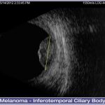
4. Melanoma – Ciliary Body
10MHz Posterior Segment B-Scan: Left ciliary body melanoma with cystic spaces as per case study 3.
Images courtesy of Bertil Damato, PhD, FRCOphth, Royal Liverpool University Hospital, United Kingdom.
-
Eye Cubed™ features real time imaging, advanced movie mode, real-time image enhancement, and a range of self-calibrating "best fit" probes.
Customized Configuration to Best Meet Your Needs
With customized configuration of A-Scan and B-Scan modes, Eye Cubed™ covers all of your diagnostic ultrasound needs for both the posterior and anterior segments. Pre-op or post-op, A-Scan or B-Scan, retina or anterior segment: whatever your focus, Eye Cubed™ shows you more, in more detail, than any other device of its kind.
High Resolution Goes Ultra
Eye Cubed’s 40 MHz UBM mode allows you to view anterior structures such as the cornea, iris, ciliary body, crystalline and intraocular lens more clearly than ever before. Whether determining the sulcus-to-sulcus measurement for accurate ICL sizing or the angle for potential angle closure and possible YAG laser iridotomy, Eye Cubed’s 40M Hz UBM mode is the gold-standard in high-resolution ultrasound.
Essential Technology for Your Practice
Even in an era of high-tech OCT scanning and digital imaging, ultrasound is the only means to obtain a crucial view of the posterior segment when there is a dense cataract or vitreous hemorrhage in the eye – making it one of the most fundamental diagnostic tools in ophthalmology. Detection of disorders like posterior vitreous detachment (PVD) in opaque ocular media is easily achieved with B-Scan ultrasound.
Innovative Imaging
Since acquiring Sacramento-based ophthalmic ultrasound pioneers Innovative Imaging Inc. in 2006, Ellex has been working hard to provide ever-increasing value to our Eye Cubed customers – and to develop our ultrasound technology platform. With the support of personalized, clinical ultrasound applications training by expert ecographers, Eye Cubed™ offers a total solution for your practice – and for your patients.
Ophthalmic Edge
Visit the comprehensive online diagnostic teaching tool created by Yale Fisher, MD, to access lectures, monographs and cases studies covering a range of diagnostic technologies, including ultrasound and OCT." href='https://www.ophthalmic.com.sg/wp-content/uploads/2016/09/04-Melanoma-Ciliary-Body-003-lg-1024x686-e1472908566394.jpg'>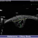
-
Eye Cubed™ features real time imaging, advanced movie mode, real-time image enhancement, and a range of self-calibrating "best fit" probes.
Customized Configuration to Best Meet Your Needs
With customized configuration of A-Scan and B-Scan modes, Eye Cubed™ covers all of your diagnostic ultrasound needs for both the posterior and anterior segments. Pre-op or post-op, A-Scan or B-Scan, retina or anterior segment: whatever your focus, Eye Cubed™ shows you more, in more detail, than any other device of its kind.
High Resolution Goes Ultra
Eye Cubed’s 40 MHz UBM mode allows you to view anterior structures such as the cornea, iris, ciliary body, crystalline and intraocular lens more clearly than ever before. Whether determining the sulcus-to-sulcus measurement for accurate ICL sizing or the angle for potential angle closure and possible YAG laser iridotomy, Eye Cubed’s 40M Hz UBM mode is the gold-standard in high-resolution ultrasound.
Essential Technology for Your Practice
Even in an era of high-tech OCT scanning and digital imaging, ultrasound is the only means to obtain a crucial view of the posterior segment when there is a dense cataract or vitreous hemorrhage in the eye – making it one of the most fundamental diagnostic tools in ophthalmology. Detection of disorders like posterior vitreous detachment (PVD) in opaque ocular media is easily achieved with B-Scan ultrasound.
Innovative Imaging
Since acquiring Sacramento-based ophthalmic ultrasound pioneers Innovative Imaging Inc. in 2006, Ellex has been working hard to provide ever-increasing value to our Eye Cubed customers – and to develop our ultrasound technology platform. With the support of personalized, clinical ultrasound applications training by expert ecographers, Eye Cubed™ offers a total solution for your practice – and for your patients.
Ophthalmic Edge
Visit the comprehensive online diagnostic teaching tool created by Yale Fisher, MD, to access lectures, monographs and cases studies covering a range of diagnostic technologies, including ultrasound and OCT." href='https://www.ophthalmic.com.sg/wp-content/uploads/2016/09/04-Melanoma-Ciliary-Body-002-lg-1024x686-e1472908552273.jpg'>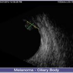
-
Eye Cubed™ features real time imaging, advanced movie mode, real-time image enhancement, and a range of self-calibrating "best fit" probes.
Customized Configuration to Best Meet Your Needs
With customized configuration of A-Scan and B-Scan modes, Eye Cubed™ covers all of your diagnostic ultrasound needs for both the posterior and anterior segments. Pre-op or post-op, A-Scan or B-Scan, retina or anterior segment: whatever your focus, Eye Cubed™ shows you more, in more detail, than any other device of its kind.
High Resolution Goes Ultra
Eye Cubed’s 40 MHz UBM mode allows you to view anterior structures such as the cornea, iris, ciliary body, crystalline and intraocular lens more clearly than ever before. Whether determining the sulcus-to-sulcus measurement for accurate ICL sizing or the angle for potential angle closure and possible YAG laser iridotomy, Eye Cubed’s 40M Hz UBM mode is the gold-standard in high-resolution ultrasound.
Essential Technology for Your Practice
Even in an era of high-tech OCT scanning and digital imaging, ultrasound is the only means to obtain a crucial view of the posterior segment when there is a dense cataract or vitreous hemorrhage in the eye – making it one of the most fundamental diagnostic tools in ophthalmology. Detection of disorders like posterior vitreous detachment (PVD) in opaque ocular media is easily achieved with B-Scan ultrasound.
Innovative Imaging
Since acquiring Sacramento-based ophthalmic ultrasound pioneers Innovative Imaging Inc. in 2006, Ellex has been working hard to provide ever-increasing value to our Eye Cubed customers – and to develop our ultrasound technology platform. With the support of personalized, clinical ultrasound applications training by expert ecographers, Eye Cubed™ offers a total solution for your practice – and for your patients.
Ophthalmic Edge
Visit the comprehensive online diagnostic teaching tool created by Yale Fisher, MD, to access lectures, monographs and cases studies covering a range of diagnostic technologies, including ultrasound and OCT." href='https://www.ophthalmic.com.sg/wp-content/uploads/2016/09/04-Melanoma-Ciliary-Body-001-lg-1024x686-e1472908538931.jpg'>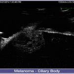
5. Melanoma – Choroid, Iris and Anterior Chamber
10MHz Posterior Segment B-Scan: A very atypical melanoma extending from choroid through iris into anterior chamber and showing necrosis.
Images courtesy of Bertil Damato, PhD, FRCOphth, Royal Liverpool University Hospital, United Kingdom.
-
Eye Cubed™ features real time imaging, advanced movie mode, real-time image enhancement, and a range of self-calibrating "best fit" probes.
Customized Configuration to Best Meet Your Needs
With customized configuration of A-Scan and B-Scan modes, Eye Cubed™ covers all of your diagnostic ultrasound needs for both the posterior and anterior segments. Pre-op or post-op, A-Scan or B-Scan, retina or anterior segment: whatever your focus, Eye Cubed™ shows you more, in more detail, than any other device of its kind.
High Resolution Goes Ultra
Eye Cubed’s 40 MHz UBM mode allows you to view anterior structures such as the cornea, iris, ciliary body, crystalline and intraocular lens more clearly than ever before. Whether determining the sulcus-to-sulcus measurement for accurate ICL sizing or the angle for potential angle closure and possible YAG laser iridotomy, Eye Cubed’s 40M Hz UBM mode is the gold-standard in high-resolution ultrasound.
Essential Technology for Your Practice
Even in an era of high-tech OCT scanning and digital imaging, ultrasound is the only means to obtain a crucial view of the posterior segment when there is a dense cataract or vitreous hemorrhage in the eye – making it one of the most fundamental diagnostic tools in ophthalmology. Detection of disorders like posterior vitreous detachment (PVD) in opaque ocular media is easily achieved with B-Scan ultrasound.
Innovative Imaging
Since acquiring Sacramento-based ophthalmic ultrasound pioneers Innovative Imaging Inc. in 2006, Ellex has been working hard to provide ever-increasing value to our Eye Cubed customers – and to develop our ultrasound technology platform. With the support of personalized, clinical ultrasound applications training by expert ecographers, Eye Cubed™ offers a total solution for your practice – and for your patients.
Ophthalmic Edge
Visit the comprehensive online diagnostic teaching tool created by Yale Fisher, MD, to access lectures, monographs and cases studies covering a range of diagnostic technologies, including ultrasound and OCT." href='https://www.ophthalmic.com.sg/wp-content/uploads/2016/09/05-Melanoma-Choroid-Iris-and-Anterior-Chamber-001-lg-1024x686-e1472908596561.jpg'>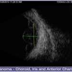
-
Eye Cubed™ features real time imaging, advanced movie mode, real-time image enhancement, and a range of self-calibrating "best fit" probes.
Customized Configuration to Best Meet Your Needs
With customized configuration of A-Scan and B-Scan modes, Eye Cubed™ covers all of your diagnostic ultrasound needs for both the posterior and anterior segments. Pre-op or post-op, A-Scan or B-Scan, retina or anterior segment: whatever your focus, Eye Cubed™ shows you more, in more detail, than any other device of its kind.
High Resolution Goes Ultra
Eye Cubed’s 40 MHz UBM mode allows you to view anterior structures such as the cornea, iris, ciliary body, crystalline and intraocular lens more clearly than ever before. Whether determining the sulcus-to-sulcus measurement for accurate ICL sizing or the angle for potential angle closure and possible YAG laser iridotomy, Eye Cubed’s 40M Hz UBM mode is the gold-standard in high-resolution ultrasound.
Essential Technology for Your Practice
Even in an era of high-tech OCT scanning and digital imaging, ultrasound is the only means to obtain a crucial view of the posterior segment when there is a dense cataract or vitreous hemorrhage in the eye – making it one of the most fundamental diagnostic tools in ophthalmology. Detection of disorders like posterior vitreous detachment (PVD) in opaque ocular media is easily achieved with B-Scan ultrasound.
Innovative Imaging
Since acquiring Sacramento-based ophthalmic ultrasound pioneers Innovative Imaging Inc. in 2006, Ellex has been working hard to provide ever-increasing value to our Eye Cubed customers – and to develop our ultrasound technology platform. With the support of personalized, clinical ultrasound applications training by expert ecographers, Eye Cubed™ offers a total solution for your practice – and for your patients.
Ophthalmic Edge
Visit the comprehensive online diagnostic teaching tool created by Yale Fisher, MD, to access lectures, monographs and cases studies covering a range of diagnostic technologies, including ultrasound and OCT." href='https://www.ophthalmic.com.sg/wp-content/uploads/2016/09/05-Melanoma-Choroid-Iris-and-Anterior-Chamber-002-lg-1024x686-e1472908585425.jpg'>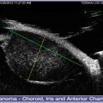
6. Melanoma – Iris
10MHz Posterior Segment B-Scan: Temporal iris melanoma.
Images courtesy of Bertil Damato, PhD, FRCOphth, Royal Liverpool University Hospital, United Kingdom.
-
Eye Cubed™ features real time imaging, advanced movie mode, real-time image enhancement, and a range of self-calibrating "best fit" probes.
Customized Configuration to Best Meet Your Needs
With customized configuration of A-Scan and B-Scan modes, Eye Cubed™ covers all of your diagnostic ultrasound needs for both the posterior and anterior segments. Pre-op or post-op, A-Scan or B-Scan, retina or anterior segment: whatever your focus, Eye Cubed™ shows you more, in more detail, than any other device of its kind.
High Resolution Goes Ultra
Eye Cubed’s 40 MHz UBM mode allows you to view anterior structures such as the cornea, iris, ciliary body, crystalline and intraocular lens more clearly than ever before. Whether determining the sulcus-to-sulcus measurement for accurate ICL sizing or the angle for potential angle closure and possible YAG laser iridotomy, Eye Cubed’s 40M Hz UBM mode is the gold-standard in high-resolution ultrasound.
Essential Technology for Your Practice
Even in an era of high-tech OCT scanning and digital imaging, ultrasound is the only means to obtain a crucial view of the posterior segment when there is a dense cataract or vitreous hemorrhage in the eye – making it one of the most fundamental diagnostic tools in ophthalmology. Detection of disorders like posterior vitreous detachment (PVD) in opaque ocular media is easily achieved with B-Scan ultrasound.
Innovative Imaging
Since acquiring Sacramento-based ophthalmic ultrasound pioneers Innovative Imaging Inc. in 2006, Ellex has been working hard to provide ever-increasing value to our Eye Cubed customers – and to develop our ultrasound technology platform. With the support of personalized, clinical ultrasound applications training by expert ecographers, Eye Cubed™ offers a total solution for your practice – and for your patients.
Ophthalmic Edge
Visit the comprehensive online diagnostic teaching tool created by Yale Fisher, MD, to access lectures, monographs and cases studies covering a range of diagnostic technologies, including ultrasound and OCT." href='https://www.ophthalmic.com.sg/wp-content/uploads/2016/09/06-Melanoma-Iris-001-lg-1024x686-e1472908638345.jpg'>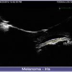
-
Eye Cubed™ features real time imaging, advanced movie mode, real-time image enhancement, and a range of self-calibrating "best fit" probes.
Customized Configuration to Best Meet Your Needs
With customized configuration of A-Scan and B-Scan modes, Eye Cubed™ covers all of your diagnostic ultrasound needs for both the posterior and anterior segments. Pre-op or post-op, A-Scan or B-Scan, retina or anterior segment: whatever your focus, Eye Cubed™ shows you more, in more detail, than any other device of its kind.
High Resolution Goes Ultra
Eye Cubed’s 40 MHz UBM mode allows you to view anterior structures such as the cornea, iris, ciliary body, crystalline and intraocular lens more clearly than ever before. Whether determining the sulcus-to-sulcus measurement for accurate ICL sizing or the angle for potential angle closure and possible YAG laser iridotomy, Eye Cubed’s 40M Hz UBM mode is the gold-standard in high-resolution ultrasound.
Essential Technology for Your Practice
Even in an era of high-tech OCT scanning and digital imaging, ultrasound is the only means to obtain a crucial view of the posterior segment when there is a dense cataract or vitreous hemorrhage in the eye – making it one of the most fundamental diagnostic tools in ophthalmology. Detection of disorders like posterior vitreous detachment (PVD) in opaque ocular media is easily achieved with B-Scan ultrasound.
Innovative Imaging
Since acquiring Sacramento-based ophthalmic ultrasound pioneers Innovative Imaging Inc. in 2006, Ellex has been working hard to provide ever-increasing value to our Eye Cubed customers – and to develop our ultrasound technology platform. With the support of personalized, clinical ultrasound applications training by expert ecographers, Eye Cubed™ offers a total solution for your practice – and for your patients.
Ophthalmic Edge
Visit the comprehensive online diagnostic teaching tool created by Yale Fisher, MD, to access lectures, monographs and cases studies covering a range of diagnostic technologies, including ultrasound and OCT." href='https://www.ophthalmic.com.sg/wp-content/uploads/2016/09/06-Melanoma-Iris-002-lg-1024x686-e1472908623104.jpg'>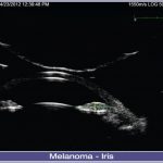
7. RPE/Choroidal Detachment
10MHz Posterior Segment B-Scan: Eccentric disciform lesion with RPE/choroidal detachment.
Image courtesy of Bertil Damato, PhD, FRCOphth, Royal Liverpool University Hospital, United Kingdom.
-
Eye Cubed™ features real time imaging, advanced movie mode, real-time image enhancement, and a range of self-calibrating "best fit" probes.
Customized Configuration to Best Meet Your Needs
With customized configuration of A-Scan and B-Scan modes, Eye Cubed™ covers all of your diagnostic ultrasound needs for both the posterior and anterior segments. Pre-op or post-op, A-Scan or B-Scan, retina or anterior segment: whatever your focus, Eye Cubed™ shows you more, in more detail, than any other device of its kind.
High Resolution Goes Ultra
Eye Cubed’s 40 MHz UBM mode allows you to view anterior structures such as the cornea, iris, ciliary body, crystalline and intraocular lens more clearly than ever before. Whether determining the sulcus-to-sulcus measurement for accurate ICL sizing or the angle for potential angle closure and possible YAG laser iridotomy, Eye Cubed’s 40M Hz UBM mode is the gold-standard in high-resolution ultrasound.
Essential Technology for Your Practice
Even in an era of high-tech OCT scanning and digital imaging, ultrasound is the only means to obtain a crucial view of the posterior segment when there is a dense cataract or vitreous hemorrhage in the eye – making it one of the most fundamental diagnostic tools in ophthalmology. Detection of disorders like posterior vitreous detachment (PVD) in opaque ocular media is easily achieved with B-Scan ultrasound.
Innovative Imaging
Since acquiring Sacramento-based ophthalmic ultrasound pioneers Innovative Imaging Inc. in 2006, Ellex has been working hard to provide ever-increasing value to our Eye Cubed customers – and to develop our ultrasound technology platform. With the support of personalized, clinical ultrasound applications training by expert ecographers, Eye Cubed™ offers a total solution for your practice – and for your patients.
Ophthalmic Edge
Visit the comprehensive online diagnostic teaching tool created by Yale Fisher, MD, to access lectures, monographs and cases studies covering a range of diagnostic technologies, including ultrasound and OCT." href='https://www.ophthalmic.com.sg/wp-content/uploads/2016/09/07-RPE-Choroidal-Detachment-001-lg-1024x686-e1472908233190.jpg'>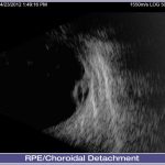
Documents
Eye Cubed Product Brochure
Download