-
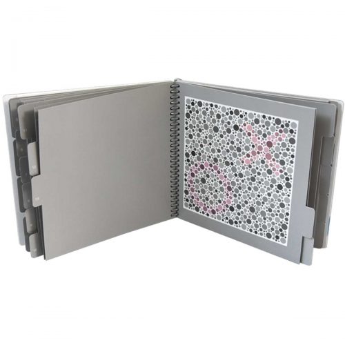 730005 The Laminated HRR (Hardy Rand and Rittler) Pseudoisochromatic Test, 4th Edition provides several very important features to provide the most advanced color vision test available: congenital and acquired testing, identification of the type of defect, and diagnosis of the extent of the defect as well as quick positive classification of normals. The HRR employs a sophisticated test strategy that virtually eliminates the potential for memorization and malingering. The figures used by the HRR Pseudoisochromatic Plates are independent of language and suitable for both adults and children.
 -
730006 The HRR (Hardy Rand and Rittler) Standard Pseudoisochromatic Test, 4th Edition provides several very important features to provide the most advanced color vision test available: congenital and acquired testing, identification of the type of defect, and diagnosis of the extent of the defect as well as quick positive classification of normals. The HRR employs a sophisticated test strategy that virtually eliminates the potential for memorization and malingering. The figures used by the HRR Pseudoisochromatic Plates are independent of language and suitable for both adults and children.
-

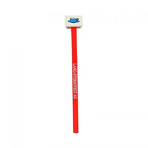
The Lang Fixation Cubes have been designed by J. Lang for the assessment of eye fixation, motility, accommodation, convergence, for cover test, and to prepare the child for testing with the Lang-Stereotest® or the Lang-Stereopad®.
5 child-friendly objects are presented on the side and the top of the cube. Some of them are also included in the two versions of the Lang-Stereotest® and of the Lang-Stereopad®, and on the Lang Fixation stick.
The Cube has a red handle.
Each turn of the cube shows a new object. The star on the top side and the other objects can also be used for the test of convergence.
-
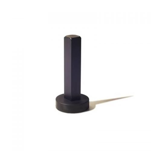 The magnetic monopod support has been designed specially to be used with the Lang-Stereopad®. The test plate can be easily placed in reading position on a table, both in portrait and landscape position. The magnetic monopod serves also as a handle on the back side of the test plate when the test needs to be shown in frontal position.
 -

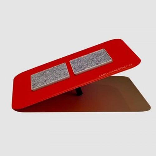
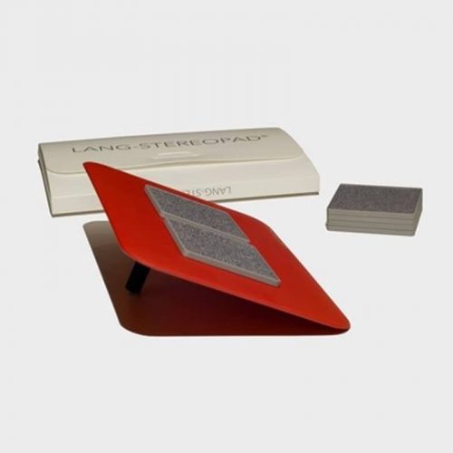 The Lang-Stereopad® is a new device for testing global near stereopsis. It is suitable for all age groups, including preverbal children (less than 1 year). This stereo test does not require the use of special glasses because of the combination of random dots with a lenticular screen, similarly to the the Lang-Stereotests® I-R and II.
 -
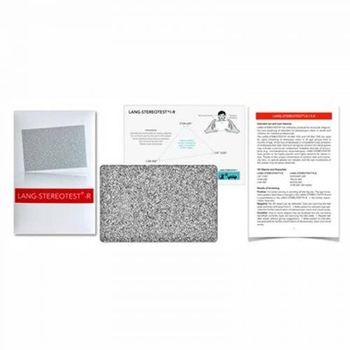
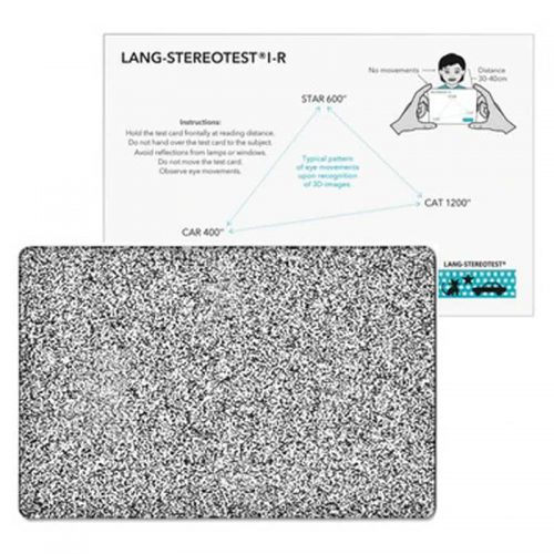 The Lang-Stereotest® I-R is an easy-to-use screening-test designed for early detection of problems with stereoscopic vision of children. In contrast to most stereo tests it does not require glasses, due to the combination of random dots and a lenticular screen.
 -
 PART #VDGTLCM The digital clear mag is designed specifically for high magnification and detailed examination of the macula and optic disc with a 3.89x magnification. This lens is an easy transition for those who are used to handling a 20D lens with a similar lens ring size and working distance but with an added advantage of high magnification. This lens is a great choice for detecting and monitoring the subtle changes in the optic disc morphology in patients with diabetic retinopathy, glaucoma and age related macular degeneration.
 -
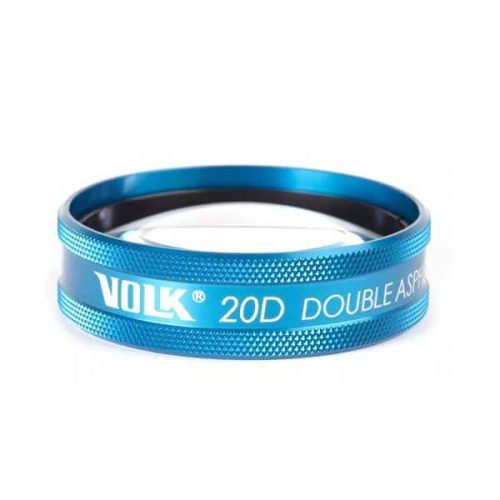
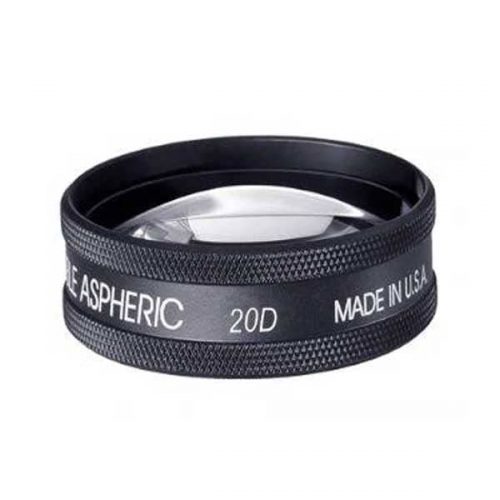 PART #V20LC Perhaps the most recognized lens around the world, the Volk 20D is the lens that started the legacy of double aspheric lens design for BIO and is still considered as a gold standard. The contributing factors for this are the perfect balance of field of view and magnification of the 20D. The working distance of 50 mm also makes lens manipulation a comfortable experience for the examiner. This lens is perfect for general diagnosis of patients with a 3x magnification and field of view that allows visualization up to the mid peripheral region. The dynamic examination with the appropriate eye movements by the patients allows viewing of the peripheral retina as well as detailed examination by BIO in the primary position provides a comprehensive estimate of retinal health. Thus, this lens is a great choice both as a first line of diagnosis as well as a high level central retinal examination.
 -
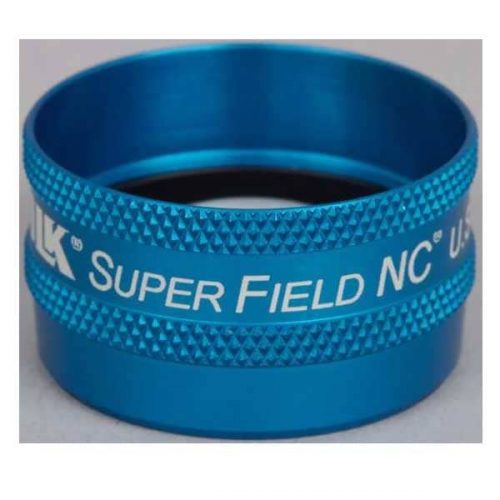
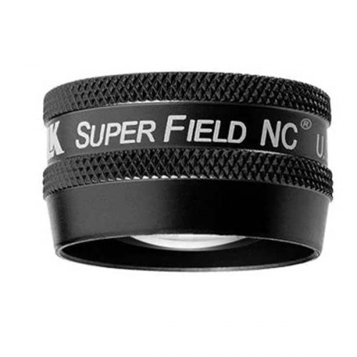 PART #VSFNC Often referred to as a ‘Super 90D’ this lens provides wide field imaging out to the mid periphery and dynamic viewing out to the periphery. This lens is a great choice for pan-retinal examination. This lens provides the magnification of a 90D, with an increased field of view enabling high resolution imaging of the posterior pole. This combination of magnification and wide field imaging allows quick and comprehensive examination of the retina, making it a go-to lens for general examination. A 30 mm lens ring provides a comfortable grip and manipulation of the lens within the orbit.
 -
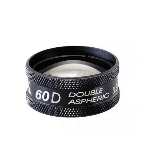 PART #V60C The 60D lens is one of the high magnification fundoscopy lenses used for a thorough and detailed examination of the central retina such as the macula and the nerve head. The trademark double aspheric design provides excellent detail and imaging for detecting subtle details and indications of retinal abnormalities. The optical profile of this lens requires a longer working distance of 18 mm from the patient. The high magnification provided by this lens is a great choice for diagnosis and assessment of the severity of Age-Related Macular Degeneration, following cup to disk ratios in patients and capillary hemorrhages. Dilation is required to obtain optimum retinal imaging with the 60D.
 -
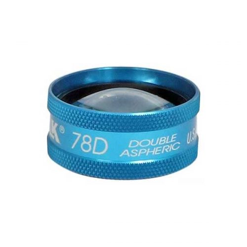
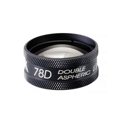 PART #V78LC The is an ideal lens for doctors who regularly cater to populations prone to glaucoma and other posterior pole abnormalities. The double aspheric design and field of view offered by a 78D offers clear and large views of the central mid-retinal regions. This lens offers a high magnification without cutting down too drastically on the field of view. This lens is a popular choice as a general diagnosis lens among doctors who prefer a larger ring size than the more common and smaller profile of the 90D. Dilation is required to obtain optimum retinal imaging with the 78D.
 -
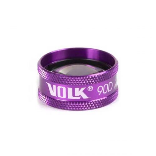
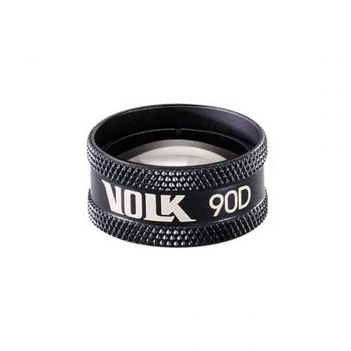 PART #V90C The Volk 90D is the most widely known fundoscopy lens and is to date considered the gold standard in exam rooms across the world. The small profile of the 90D coupled with its optical profile makes it a great first lens. This lens is a perfect choice for general examination and retinal imaging. This lens can be used to get through small pupils for patients who do not prefer or accommodate dilation and as a result is also a popular choice for undilated retinal exams.
 -
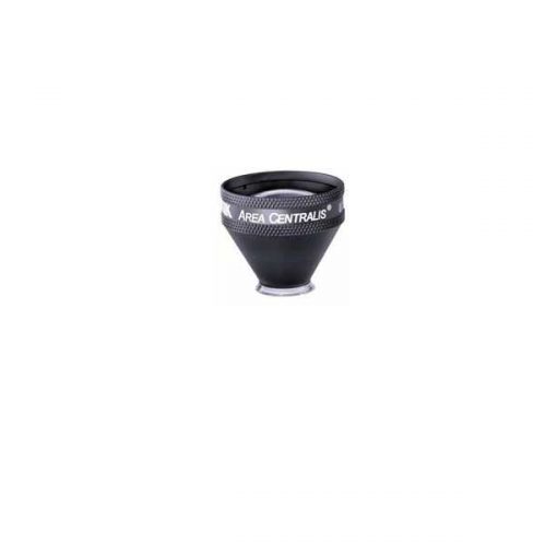 PART #VAC The Area centralis is designed with a 1.06x magnification to provide high detail, magnified views of the posterior pole. This lens is ideal for grid/focal laser procedures of the central retina for treating microaneurysms and edema in conditions such as diabetic retinopathy.
- High magnification for detailed examination of the posterior pole
- Available in flange, no flange and ANF+ contact options
- Ideal for focal/grid laser therapy
-
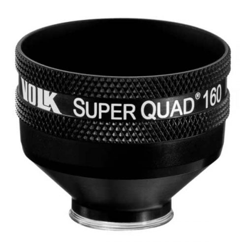 PART #VSQUAD160 Experience wide field, distortion-free visualization of the retina from the nerve head and macula, up to the ora serrata, designed for detection and treatment of retinal abnormalities like peripheral retinal tears, peripheral retinal detachments, giant retinal tears etc. The 30 mm lens surface offers a large, clear image of the retina for accurate and easy placement of the laser spot. The contact surface is designed carefully to provide optimum stability on the patient’s cornea while ensuring patient comfort.
- Wide-field distortion free viewing
- Ideal for detecting and treating mid to far-peripheral retinal abnormalities
- Large lens surface area providing a large working area
- Available in: Flanged contact, no Flange contact design
-
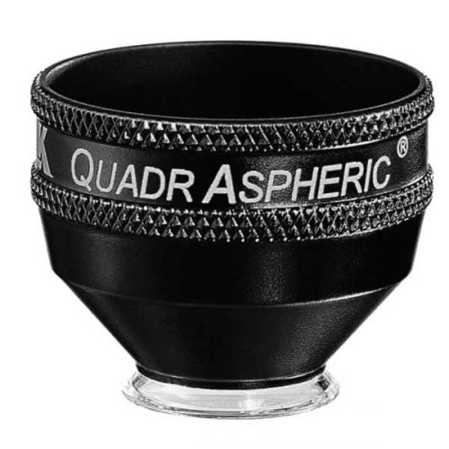 With a 144° field of view, this lens enables wide field visualization up to the peripheral retina for diagnosis and treatment of peripheral retinal defects. This lens is specially designed to provide wide field visualization even through small pupils. This application is critical when evaluating and treating patients such as those at risk of angle closure, neovascularization of the iris etc. in whom dilation should be avoided. The small pupil capability is also advantageous when treating geriatric populations in whom pupil response to dilation is limited. The large flange on this lens provides the perfect stability and control over the eye needed during laser procedures
- Wide-field distortion free viewing of the retina
- Small-pupil capability
- Ideal for detecting and treating mid to peripheral retinal abnormalities
- Available in Flange, no Flange and ANF+ contact options
-
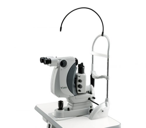
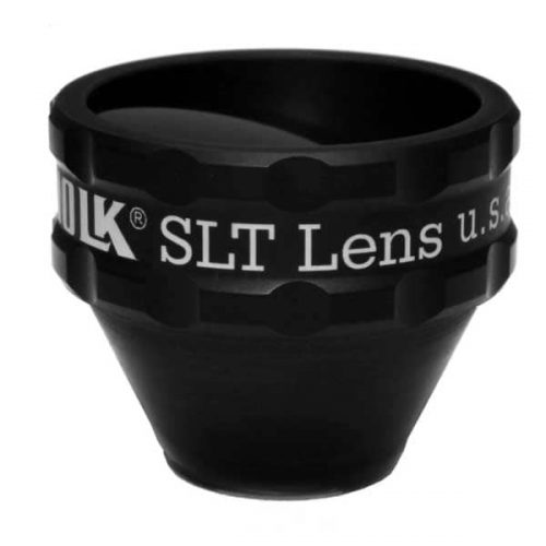 PART #VSLT This lens is a classic one mirror design lens that provides a large, clear view of the angle structures for performing SLT procedures to lower intraocular pressure in patients with ocular hypertension and glaucoma. This lens is designed for compatibility when used with a frequency doubled Q switched Nd:YAG laser. The lens has to be rotated gently on the patient’s eye to aim the laser at the various sections of iridocorneal angle. The 1.0x magnification of this lens helps maintain the laser spot size and density during laser delivery.
- Selective Laser Trabeculoplasty (SLT)
- Ideal for Glaucoma Treatment
- Laser Trabecular Meshwork treatment
-
 Silverstone, the most powerful tool yet for examining the retina, is the only ultra-widefield imaging device with integrated swept source OCT. Silverstone produces a 200° single-capture retinal image of unrivaled clarity in less than ½ second and enables optomap guided OCT scanning across the retina and into the far periphery.
-
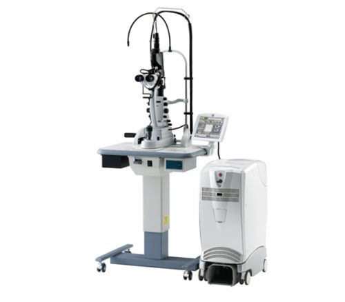
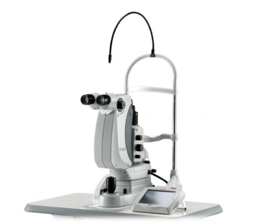 Features
- Refined laser delivery with lower energy
- SLT mode
- Clear and sharp field of view
- Precise aiming beam
- Optimized operating distance
- Unique joystick
-
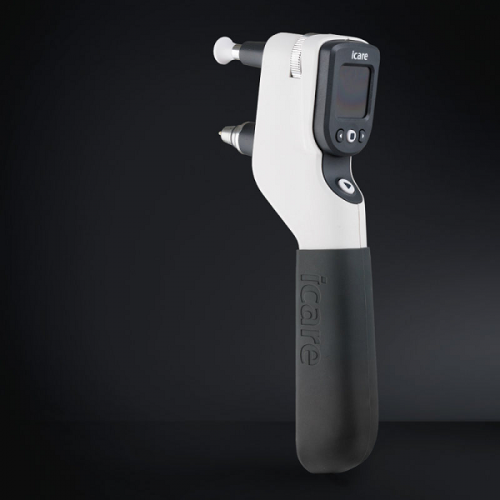
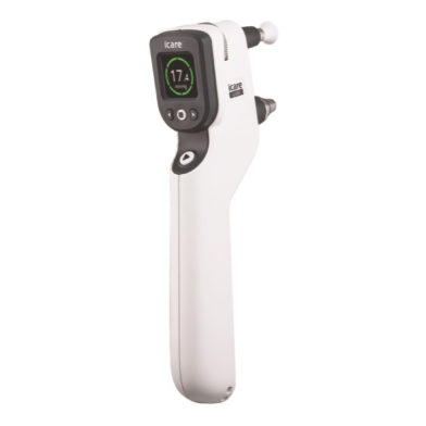
iCare IC200 tonometer - introducing a new era in clinical tonometry
Key features
- 200-degree position freedom
- Suitable for all patients
- Consistent and accurate readings
- No anesthetic drops
- Improved probe control
- User interface in multiple languages
- Wireless connection to iCare EXPORT
- Wireless printing
- No calibration
-
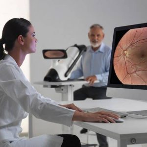
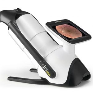
iCare DRSplus TrueColor confocal fundus imaging system
Key features- TrueColor Confocal Technology
- Multiple imaging modalities including red-free, external eye and stereo view imaging
- 2.5 mm minimum pupil size
- Fast, easy and fully automated operations
- Mosaic function which creates retinal panoramic views up to 80°
- Remote Viewer that allows for reviewing from devices on the same local area network
- Remote Exam feature enables executing an exam from a distance
-
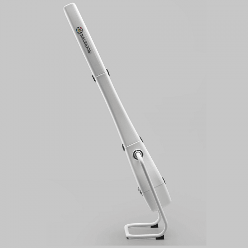
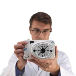
 The Kaleidos is a binocular mobile refractometer and vision analyzer that measures the refraction of both eyes and discovers other ocular impairments. The device serves as a darkroom and allows the exam to be performed in any light condition: while the patient looks inside of it, the system automatically detects refractive errors in less than three seconds. The Kaleidos measures objective refraction in the range of -15D to +15D, and phorias/tropias in prismatic diopters, as well as other additional features.
 -
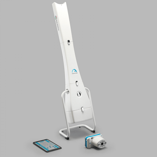
 The 2WIN is a mobile binocular video refractometer and vision analyzer that measures both eyes at the same time, in real life vision conditions. It embodies the best and the most complete technologies to fully detect refractive errors, eye abnormalities and vision problems. It measures in the range of -15D to +15D for automatic measurement of dynamic pupils response to programmable light stimulations, and accurately center spectacle lenses.
 -
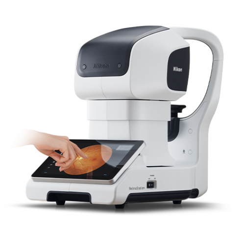
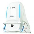
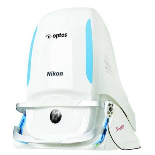
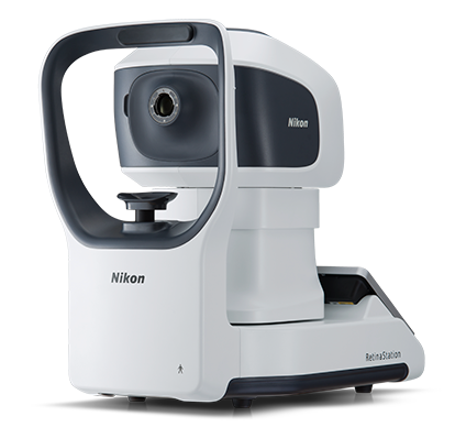 HIGHLY EFFECTIVE RETINAL EXAM The RetinaStation system is a fully-automatic, non-mydriatic 45° retinal imaging device with an eye to the 3 smart features. Retinal examination has been proven useful in diabetic retinal disease, cardiovascular disorder and other clinical cases. Nikon has taken additional steps to enhance such applications. Under its retinal imaging project, Nikon is working on brand-new retinal imaging devices.
 -
 Features
- Multicolor on modular architecture
- Multiple scan patterns
- Auto forward
- LPM (Low Power Mode)
- Practical and user-friendly features
- Delivery unit options
-
 Features
- Providing a comprehensive solution for retina and glaucoma analysis
- Accurate image capture with a SLO-based eye tracer
- Selectable OCT sensitivity that allows acquisition of B-scan images through media opacities
- Tracing HD for accurate averaging of up to 120 image
- Glaucoma analysis with wide-area normative database 9 x 9 mm
- High resolution AngioScan OCT-Angiography image
-
 Features
- The ultimate multimodal imaging platform For the SLO/OCT model - Color / FA / ICG / Blue-FAF / Green-FAF / Retro mode - OCT / OCT-Angiography* For the SLO model - Color / FA* / ICG* / Blue-FAF /Green-FAF / Retro mode
- Ultra wide field x ultra HD image*
- Unsurpassed color
- Dynamic/Simultaneous FA and ICG
- Unique Retro mode
- HD wide area OCT
- Fly Through function* Optional
-
 Beyond Compare - Like Nothing Else -
- Distortion check
- Green light transmittance measurement
- Automatic lens type detection
- Green measurement light
- A new lens table expanding measurement range
- PD measurement (only for the LM-1800PD)
- Prism layout function
- Improved marking dots
- UV transmittance measurement
- High-speed line printer with auto cutter
- Full graphic LCD with 5.7-inch touch color panel
- User-friendly tiltable LCD
- Built-in Eye Care card system
- Interface enhancement
- Refractive index measurement
-
 Features
- Advanced operation
- Accurate measurement
- Flexible and space-saving design
- Practical and user-friendly features
- Network configuration with high flexibility
-
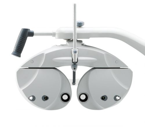
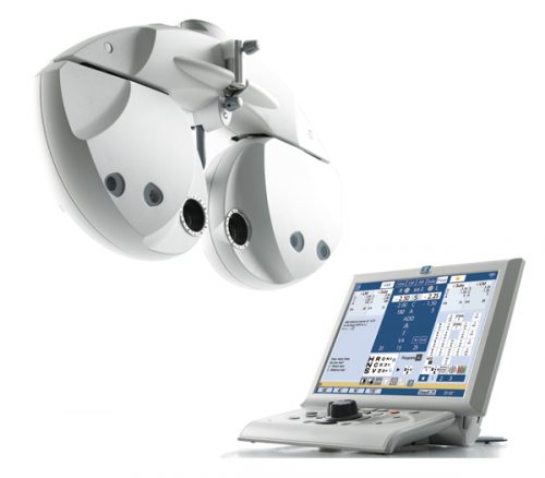 Features
- Breakthrough refractor for precise and efficient examinations
- Streamlined refractor head
- User-friendly control console
- Binocular open refraction
- Program edit function
- Simplified data transfer
-
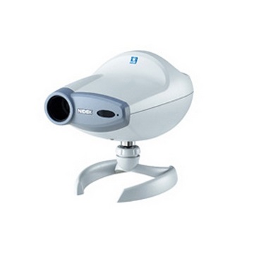
 Features
- Long-life light source for improved cost efficiency
- Brighter and clearer chart display with white LED
- Optical zoom for a variable installation distance
- Sophisticated design
- Easy focus adjustment by rotational LED aperture (available for the CP-9 EF)
-
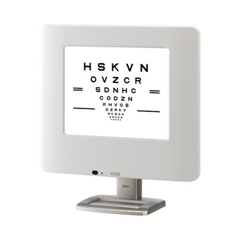 In contrast to conventional visual acuity systems, the SC-1600 displays the digitally stored chart images directly on the LCD screen
- Selectable Working Distance
- Wide Range of Image Dispkay
- Contrast Test
- Night Mode
- Indication of a Slide Image
- Black and White Inverse Function
- Smart Display Functions
-
 Key Features
- Long-life light for improved cost efficiency
- Brighter and clearer chart display with white LED
- Optical zoomer fo a variable installation distance
- Sophisticated design
-
- Minimum 0.9 m installation distance
- Combined with the Optometry System
- Night mode
- Operation with remote control
-
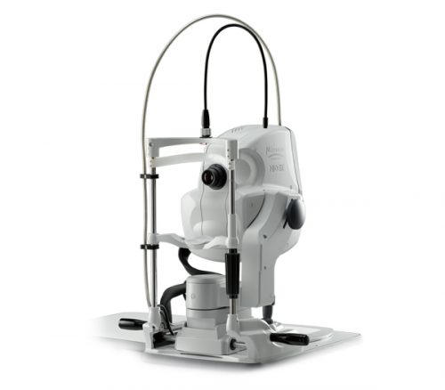
- Softer and Quiter Air Puff
- 3-D Auto Tracking, Auto Shot, and Auto Complete
- IOP Compensation by Corneal Thickness
- Attractive Tiltable 5.7-inch Color LCD
- Motorized Chinrest
- Patient Safety
- Printer with Easy Loading & Auto Detachment
-
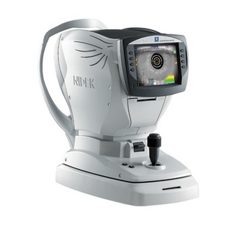
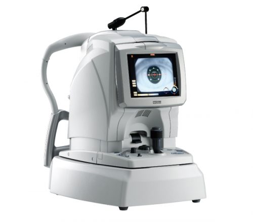
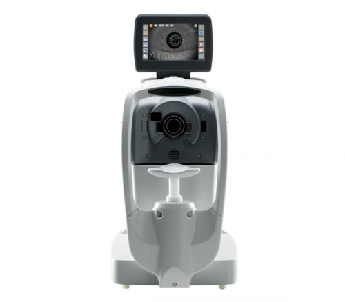
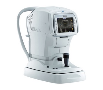
Auto Ref / Keratometer
ARK-1s / 1a / 1
- Accurate refraction measurement
- VA measurement with glare test (available for the ARK-1s)
- Simple opacity assessment with retroillumination image (available for the ARK-1s / 1a)
- Patient-friendly accommodation measurement (available for the ARK-1s / 1a)
- Keratometry measurement with mire ring
-
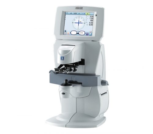
 Intuitive Design and Refined Accuracy
- Stability as Standard
- Binocular / Monocular PD Measurement
- More Precise Hairline Setting
- Auto PD Calculation with One Measurement
- Original Coated Measuring Window
- Stable Grip Design
- Digital Display
+65 6514 0848|info@ophthalmic.com.sg