Item #: BC1270SET
-
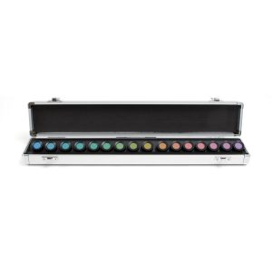
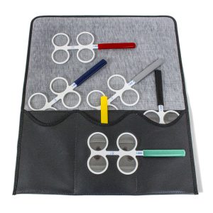 FLIPPER CASE INCLUDED. Professional level six piece set features plus/minus powers in 0.25, 0.50, 1.00, 1.50, 2.00 and 2.50 diopters. Deluxe black carrying case or the economy flipper holder included. Specify which you prefer. Flippers come with colored handle covers. Your account must be registered as a healthcare professional or a school/government in order to purchase this product.
 -
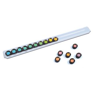
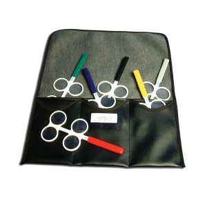 Flippers are used to test and train for accomodative facility, accommadative rock, positive/negative relative accommodation and convergence and fusional convergence reserves. May delay bifocal use with some early presbyopes. Flippers assists in hyperopia screening and lens demonstrations. Help determine dominancy and powers when fitting monovision contacts. Durable white plastic holds shape under low heat for lens insertion. Powers listed on handle. Deluxe Black carrying case or the economy flipper holder included. Item #: BC1270SETSP
 -
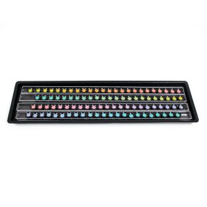
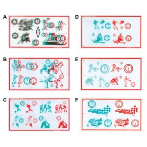 Fixed Demands Tranaglyph™ Kit (50 Series) Ideal for testing or training in true space at the office or home. Eliminates suppression while developing fusion and stereopsis. Non-variable prismatic Red/Green slides build fusional convergence & divergence. 2PD increments from 0 to 30PD BI or BO. 10" x 5.5" Red/Green targets on frosted vinyl. Can be used with Polachrome Trainer.Item #: BC50K+
 -
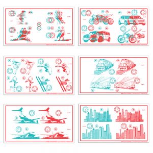 Fixed Demands Tranaglyph™ Kit (60 Series) Ideal for testing or training in true space at the office or home. Eliminates suppression while developing fusion and stereopsis. Non-variable prismatic Red/Green slides build fusional convergence & divergence. 2PD increments from 0 to 30PD BI or BO. 10" x 5.5" Red/Green targets on frosted vinyl. Can be used with Polachrome Trainer. Item #: BC60K+
 -
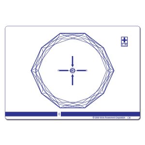
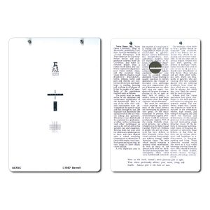 Variable slide adjust to determine fixation disparity or may be used with hand held prisms. Designed for phoroptor use. The card comes with a label which may be placed over the prism diopter scale for an additional scale in degrees. Reverse side of card includes reduced Snellen, fusional target and cross cylinder targets. 5-1/2" x 8" card and polarized goggle. Item #: BCFDC
 -
 Farnsworth Test for Congenital and Acquired color defects. The Farnsworth D-15 contains a reference disc holding notation 10 B 5 4 and fifteen numbered discs which make up an incomplete color circle. The patient arranges the discs and then evaluation of the patients arrangement separates 'normal' color perception from moderate and strong defects in deutan, protan or tritan axis color discrimination. The disks are spread out on a table and arranged by the patient. The Farnsworth D-15 test is a subset of the well known Farnsworth 100 Hue Test. It is intended for classification instead of in-depth study of color vision defects. The D-15 and 100 hue tests are correlated. Growing Importance of Color deficiency Screening: In addition to congenital color deficiency screening there is growing evidence that adult acquired color deficiency, especially in yellow and blue perception, can indicate medical toxicity and other problems. Increasingly complex security and medical systems also require verification of all three types of color receptors. How the D-15 test works: The Farnsworth D15 is called 'dichotomous' because it is designed to separate subjects into one of two groups, 1) Strongly color deficient or 2) Mildly color deficient or color normal. This is accomplished by the arrangement of vivid (saturated) colored discs. A perfect score shows normal color perception. A non-perfect score is used to determine a medium or strong color deficiency.
 -
 Farnsworth D15 Dichotomous Color Test Farnsworth Test for Congenital and Acquired color defects. The Farnsworth D-15 contains a reference disc holding notation 10 B 5 4 and fifteen numbered discs which make up an incomplete color circle. The patient arranges the discs and then evaluation of the patients arrangement separates 'normal' color perception from moderate and strong defects in deutan, protan or tritan axis color discrimination. The D-15 is housed in a plexiglass container. the disks are spread out on a table and arranged by the patient. The Farnsworth D-15 test is a subset of the well known Farnsworth 100 Hue Test. It is intended for classification instead of in-depth study of color vision defects. The D-15 and 100 hue tests are correlated. Growing Importance of Color deficiency Screening. In addition to congenital color deficiency screening there is growing evidence that adult acquired color deficiency, especially in yellow and blue perception, can indicate medical toxicity and other problems. Increasingly complex security and medical systems also require verification of all three types of color receptors. How the D-15 test works: The Farnsworth D15 is called 'dichotomous' because it is designed to separate subjects into one of two groups, 1) Strongly color deficient or 2) Mildly color deficient or color normal. This is accomplished by the arrangement of vivid (saturated) colored discs. A perfect score shows normal color perception. A non-perfect score is used to determine a medium or strong color deficiency.
 -
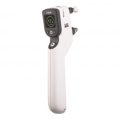
 This handheld circular pupillometer device is used for measuring different size pupils. It was developed and researched by Dr. Jack Richman, O.D., as part of the Massachusetts Drug Evaluation and Classification Program (MDEP). This pupil measurement device is used by law enforcement officers across the country to assist in determining whether people are possibly impaired and/or under the influence of drugs. The reverse side has expected pupil size in different lighting conditions for non-impaired people. Item #: LV3553000
 -
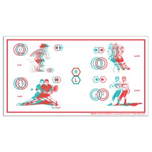
 Gem Polarized Variable Vectographic with Fixation Disparity Target → Stereopsis depth of 700 seconds of arc → Includes Fixation Disparity target with central Fusion Lock → 16 diopter range for Base-In (Divergence) Training → 24 diopter range for Base-Out (Convergence) Training → Total accommodation range of 40 diopters → New improved Vecto Guides → Now includes Therapy Binder → Now includes doctor and patient instruction manuals → Now includes Patient Vision Therapy Record Form → Includes Standard Polarized Viewers Item #: VA1060PL
 -
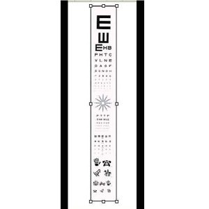 Projector Slide Replacements Family Practice Slide Letter Acuity, Astigmatic Clock, Tumbling "E", Allen Figures This family of projector slides are designed for testing all ages. Made of high quality photographic film, sealed between two pieces of glass to ensure sharp optotypes to maintain the integrity of your exam. Each slide must be adjusted for use on different length testing lanes.
 -
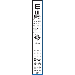 Designed for use in manual Chart Projectors including products from: R.H. Burton, Marco, Reichart, Topcon, and Woodlyn. Includes test for: → Tumbling E 20/400 – 20/200 (3 Lines) → Sloan Letters 20/100 – 20/10 (11 Lines) → Astigmatic Clock → Sloan Letters 20/50 – 20/10 (6 Lines) → Tumbling E 20/50 – 20/15 (5 Lines) → Allen Acuity 20/100 – 20/40 (4 Lines) Item #: VA1193
 -
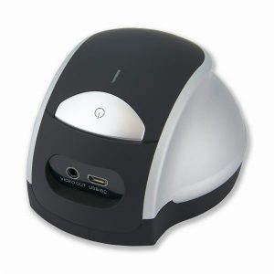
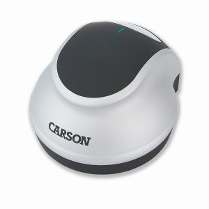 The DR-300 ezRead™ from Carson Optical transforms your television into a visual aid! Place the ezRead™ directly on top of your reading material to display the magnified image in full color on your television screen! Connect to your television in seconds using the supplied video cable. Internal led illumination ensures that the image on the screen is bright and easy to read. The size of your television monitor determines the actual magnification of the item. The larger the monitor, the greater the ezRead™ magnifies! The ezRead™ is ideal for those with low vision or macular degeneration. Operates with three AAA batteries (not included) or supplied A/C power adaptor. Magnification Table: 7x on a 17″ monitor, 11.5x on a 27″ monitor, 13.5x on a 32″ monitor, 17x on a 40″ monitor & 19.5x on a 46″ monitor