- High magnification for detailed examination of the posterior pole
- Available in flange, no flange and ANF+ contact options
- Ideal for focal/grid laser therapy
-
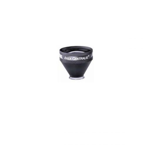 PART #VAC The Area centralis is designed with a 1.06x magnification to provide high detail, magnified views of the posterior pole. This lens is ideal for grid/focal laser procedures of the central retina for treating microaneurysms and edema in conditions such as diabetic retinopathy.
 -
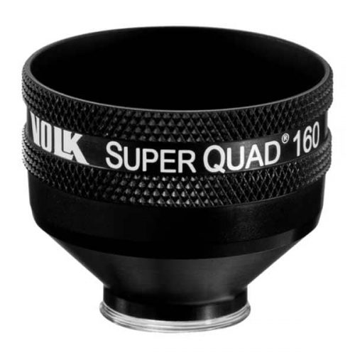 PART #VSQUAD160 Experience wide field, distortion-free visualization of the retina from the nerve head and macula, up to the ora serrata, designed for detection and treatment of retinal abnormalities like peripheral retinal tears, peripheral retinal detachments, giant retinal tears etc. The 30 mm lens surface offers a large, clear image of the retina for accurate and easy placement of the laser spot. The contact surface is designed carefully to provide optimum stability on the patient’s cornea while ensuring patient comfort.
- Wide-field distortion free viewing
- Ideal for detecting and treating mid to far-peripheral retinal abnormalities
- Large lens surface area providing a large working area
- Available in: Flanged contact, no Flange contact design
-
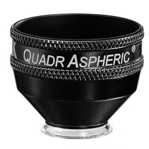 With a 144° field of view, this lens enables wide field visualization up to the peripheral retina for diagnosis and treatment of peripheral retinal defects. This lens is specially designed to provide wide field visualization even through small pupils. This application is critical when evaluating and treating patients such as those at risk of angle closure, neovascularization of the iris etc. in whom dilation should be avoided. The small pupil capability is also advantageous when treating geriatric populations in whom pupil response to dilation is limited. The large flange on this lens provides the perfect stability and control over the eye needed during laser procedures
- Wide-field distortion free viewing of the retina
- Small-pupil capability
- Ideal for detecting and treating mid to peripheral retinal abnormalities
- Available in Flange, no Flange and ANF+ contact options
-
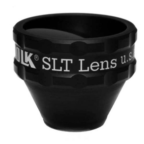 PART #VSLT This lens is a classic one mirror design lens that provides a large, clear view of the angle structures for performing SLT procedures to lower intraocular pressure in patients with ocular hypertension and glaucoma. This lens is designed for compatibility when used with a frequency doubled Q switched Nd:YAG laser. The lens has to be rotated gently on the patient’s eye to aim the laser at the various sections of iridocorneal angle. The 1.0x magnification of this lens helps maintain the laser spot size and density during laser delivery.
- Selective Laser Trabeculoplasty (SLT)
- Ideal for Glaucoma Treatment
- Laser Trabecular Meshwork treatment
-
 Silverstone, the most powerful tool yet for examining the retina, is the only ultra-widefield imaging device with integrated swept source OCT. Silverstone produces a 200° single-capture retinal image of unrivaled clarity in less than ½ second and enables optomap guided OCT scanning across the retina and into the far periphery.
-
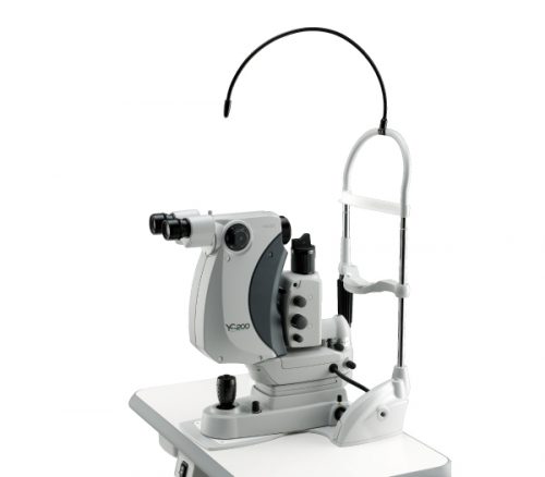
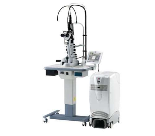
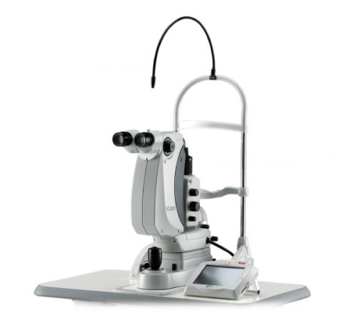 Features
- Refined laser delivery with lower energy
- SLT mode
- Clear and sharp field of view
- Precise aiming beam
- Optimized operating distance
- Unique joystick
-
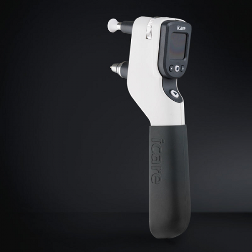
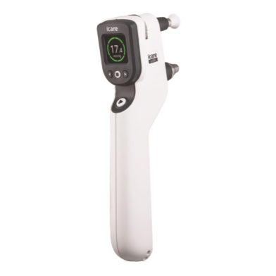
iCare IC200 tonometer - introducing a new era in clinical tonometry
Key features
- 200-degree position freedom
- Suitable for all patients
- Consistent and accurate readings
- No anesthetic drops
- Improved probe control
- User interface in multiple languages
- Wireless connection to iCare EXPORT
- Wireless printing
- No calibration
-

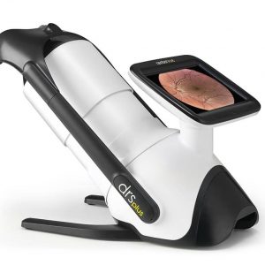
iCare DRSplus TrueColor confocal fundus imaging system
Key features- TrueColor Confocal Technology
- Multiple imaging modalities including red-free, external eye and stereo view imaging
- 2.5 mm minimum pupil size
- Fast, easy and fully automated operations
- Mosaic function which creates retinal panoramic views up to 80°
- Remote Viewer that allows for reviewing from devices on the same local area network
- Remote Exam feature enables executing an exam from a distance
-
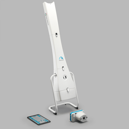 The Kaleidos is a binocular mobile refractometer and vision analyzer that measures the refraction of both eyes and discovers other ocular impairments. The device serves as a darkroom and allows the exam to be performed in any light condition: while the patient looks inside of it, the system automatically detects refractive errors in less than three seconds. The Kaleidos measures objective refraction in the range of -15D to +15D, and phorias/tropias in prismatic diopters, as well as other additional features.
 -
 The 2WIN is a mobile binocular video refractometer and vision analyzer that measures both eyes at the same time, in real life vision conditions. It embodies the best and the most complete technologies to fully detect refractive errors, eye abnormalities and vision problems. It measures in the range of -15D to +15D for automatic measurement of dynamic pupils response to programmable light stimulations, and accurately center spectacle lenses.
 -
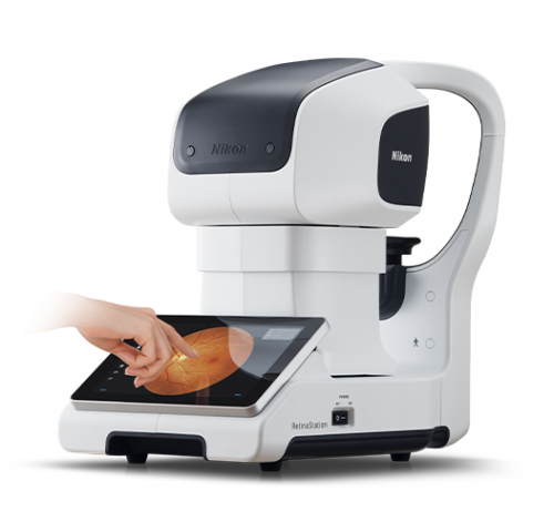
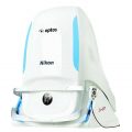
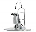
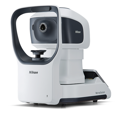 HIGHLY EFFECTIVE RETINAL EXAM The RetinaStation system is a fully-automatic, non-mydriatic 45° retinal imaging device with an eye to the 3 smart features. Retinal examination has been proven useful in diabetic retinal disease, cardiovascular disorder and other clinical cases. Nikon has taken additional steps to enhance such applications. Under its retinal imaging project, Nikon is working on brand-new retinal imaging devices.
 -
 Features
- Multicolor on modular architecture
- Multiple scan patterns
- Auto forward
- LPM (Low Power Mode)
- Practical and user-friendly features
- Delivery unit options