-
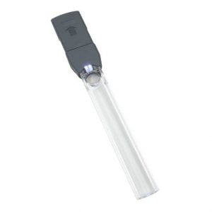
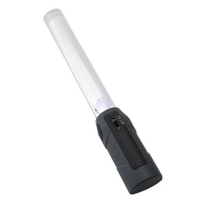 Carson’s MagniBar™ is a 2x bar magnifier with a spot lens that magnifies up to 12x. The dual LED lights feature 3 brightness settings. Great for small print reading such as newspapers, magazines and directories. US Patent D675,659
 -
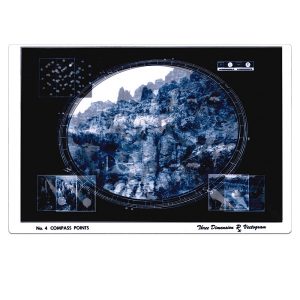
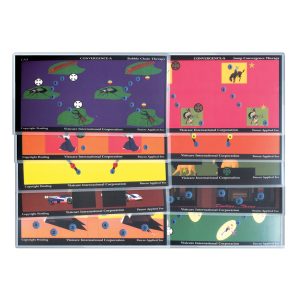 Morgenstern Color Fusion Cards - Convergence A Dr. Only Used with Bernell-O-Scope™ CONVERGENCE A. TRADITIONAL VT FOR FUSION RANGES & JUMP DUCTIONS WITH COLORFUL TARGETS. INCREASE IN DEMAND AND RANGE OF FLEXIBILITY. Item #: SMCA
 -
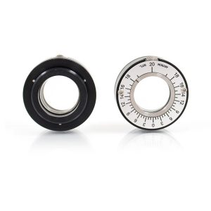 Fits into the standard 38 mm eyewall of the Bernell-O-Scope™ or and standard trial lens frame. Each prism yields 20 PD for a total of 40PD. Now you can set any amount of BI, BO, or vertical prism into the stereoscope to do training procedures. May be used for distance training by removing the cardholder. It may also be used with the new Bernell 3-D videos. Item #: ARPHH20S
 -
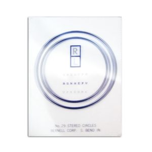 Improve vertical or horiontal fusional reserves. Dispense to patients wearing vertical prism or with low fusional reserves. Does not fit in Slide Illuminators. Used with the vertical mini-vecto kit (BC275MVK) and with the horizontal Mini-Vecto Kit(BC216MVK) Item #: BC29
 -
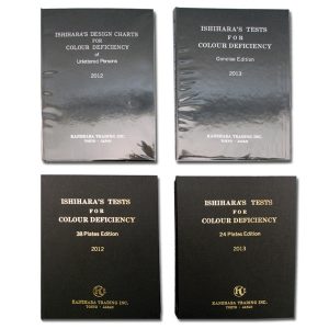
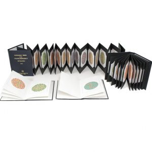 Ishihara Pseudo-Isochromatic Test The most widely used standard of color vision screening in a book form. Instructions Included.
 -
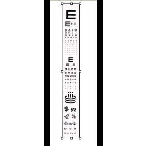 Projector Slide Replacements MultiAcuity Projector Slide Letter Acuity, Tumbling "E", Allen Figure Test This family of projector slides are designed for testing all ages. Made of high quality photographic film, sealed between two pieces of glass to ensure sharp optotypes to maintain the integrity of your exam. Each slide must be adjusted for use on different length testing lanes.
 -
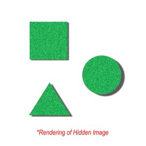
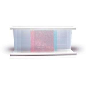 Bernell Red/Green Random Dot Variable Tranaglyph Square, Circle and Triangle Targets Now you can train using high levels of binocular skills. This slide can allow those with high amounts of esophoria or exophoira to experience random dot 3-D pictures for the first time. Use with our R/G flipper and get even more flexibility in training. (*Pictured with Sided Holder, not included)
 -
 7-1/2" x 5-1/2" Non-Variable Polarized vectograms ensure testing and training at actual distances. Vectograms stabalize fusion and steropsis, eliminate suppression and develop simultaneous vision. Used in monocular, binocular, pursuit and saccdic training, vectograms help correct and prevent anomalies involving projection and hand-eye coordination. Ideal for use with Bernells Dual Polachrome™ Trainer
 -
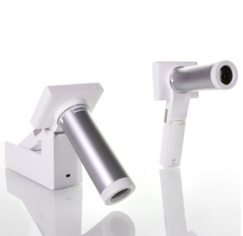
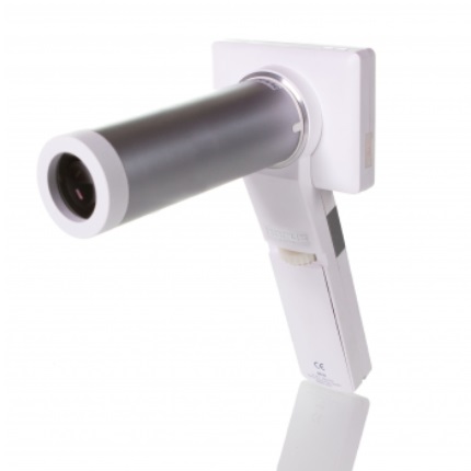
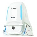
- Avoid unnecessary mydriatic medications and side effects.
- Improve quality and efficiency of making the rounds and even reach those in remote areas.
- Minimize paperwork with images or videos taken, stored, and processed digitally.
- Acquire additional stability with a special adapter attached to slit lamp.
-
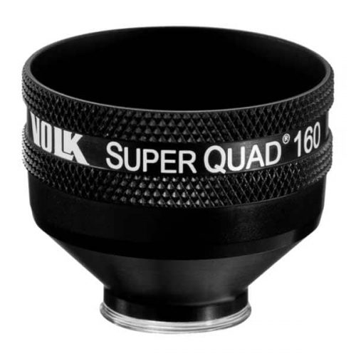 PART #VSQUAD160 Experience wide field, distortion-free visualization of the retina from the nerve head and macula, up to the ora serrata, designed for detection and treatment of retinal abnormalities like peripheral retinal tears, peripheral retinal detachments, giant retinal tears etc. The 30 mm lens surface offers a large, clear image of the retina for accurate and easy placement of the laser spot. The contact surface is designed carefully to provide optimum stability on the patient’s cornea while ensuring patient comfort.
- Wide-field distortion free viewing
- Ideal for detecting and treating mid to far-peripheral retinal abnormalities
- Large lens surface area providing a large working area
- Available in: Flanged contact, no Flange contact design
-
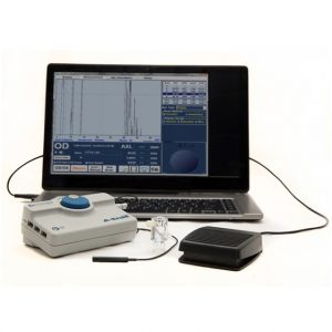
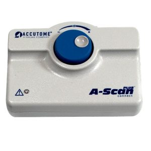 The A-Scan Plus Connect® is designed to meet the demands of today’s refractive cataract surgery. It is the single most effective solution for measuring and calculating all of your patients and refining your surgical outcomes. It has the ability to measure most patients and works great with dense cataracts or patients with fixation difficulties. The Connect links directly to a PC, laptop or tablet device making it the ultimate portable solution. Information can easily be uploaded to electronic medical record (EMR) systems. The improved user interface makes entering patient data, capturing the scan and calculating the measurement much faster.* Features:
- Immersion and Contact Modes.
- Industry Leading Resolution.
- User-friendly interface.
- Less Patient Chair Time – Faster measurement capture*.
- Automatic Alignment Detection and Sclera Recognition eliminate marginally aligned scans.
- Portable, Lightweight Design – Plug into any PC, laptop or tablet device.
- Share information easily – adaptable document transfer via EMR, email or printer.
- Modern Third and Fourth Generation Formulas including the Hoffer®Q, SRK/T, Holladay, Haigis as well as new post refractive formulas.
- Optimized Lens Constants for superior surgical outcomes.
- Unlimited Software Licenses.
- Streamline your patient data - shares information with B-Scan Plus and UBM Plus.
-
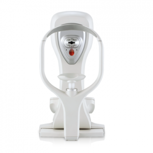
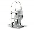
 The revolutionary OCULUS Corvis® STL records the reaction of the cornea to a defined air pulse with a newly developed high-speed Scheimpflug-camera that takes over 4,300 images per second. IOP and corneal thickness can be measured with great precision on the basis of the Scheimpflug images.
Corneal Visualization Scheimpflug Technology
The revolutionary OCULUS Corvis® STL records the reaction of the cornea to a defined air pulse with a newly developed high-speed Scheimpflug-camera that takes over 4,300 images per second. IOP and corneal thickness can be measured with great precision on the basis of the Scheimpflug images.Measurement and Display Options
- IOP measurement
- Measurement of the corneal thickness
- Scheimpflug images of the 1st and 2nd applanation of the cornea
- Slow-motion video of the corneal deformation as a result of the air pulse
- Non-contact tonometer in combination with an ultra-high-speed camera for visualization of the deformation of the cornea in reaction to an air pulse (4,330 frames / sec.)
+65 6514 0848|info@ophthalmic.com.sg