

- Avoid unnecessary mydriatic medications and side effects.
- Improve quality and efficiency of making the rounds and even reach those in remote areas.
- Minimize paperwork with images or videos taken, stored, and processed digitally.
- Acquire additional stability with a special adapter attached to slit lamp.
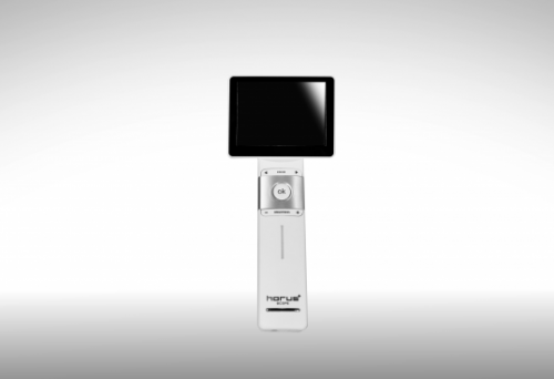
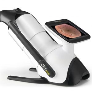
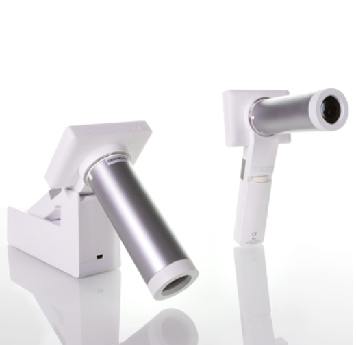
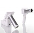
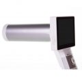
Horus Digital Fundus Camera DEC 100
Scenario
- Emergency
- Pediatrics
- Ward round
- Clinics
- Telemedicine
- Education
- Veterinary
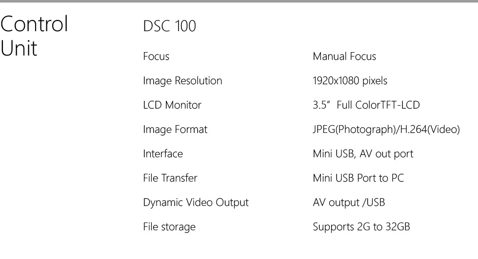
Technical Specifications
QnA
Q: Is dilation on examinee necessary before using DEC 100? What is the minimum pupil size that allows DEC 100 to operate?
A: No, dilation is not necessary. Horus eye-fundus camera can successfully capture images on pupils larger than 3.2mm with proper light conditions. Strongly recommended to operate Horus eye-fundus camera in darkroom to obtain best quality images, or use eye cup to reduce the interference of external light source.
Q: Can Horus eye-fundus camera be used to capture images of the other segments of an eye? E.g. anterior of an eye?
A: This eye-fundus camera is designed to capture eye-fundus images only, not suitable to other usages. For devices made to capture eye anterior images, please refer to Product 6.6 Gonioscope
Q: Adjust the brightness of eye-fundus camera
A: As the level of pigmentation of people differs from each other, the brightness of eye-fundus camera should be adjusted based on examinee’s condition. The rule of thumb is to adjust the brightness to 6B for Caucasians, 8B to 10B for Asians, and 12B for Hispanics, Indians and Africans.
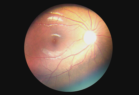
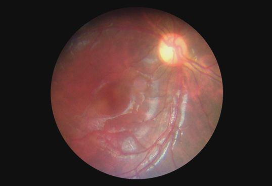
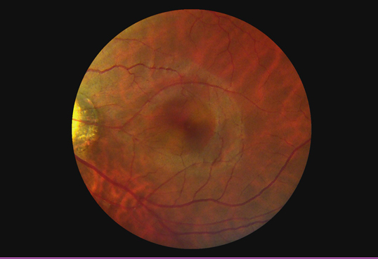
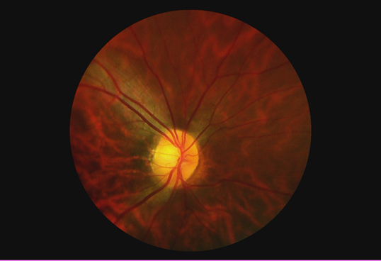
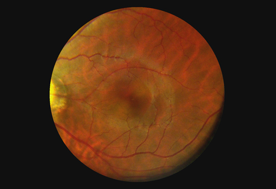
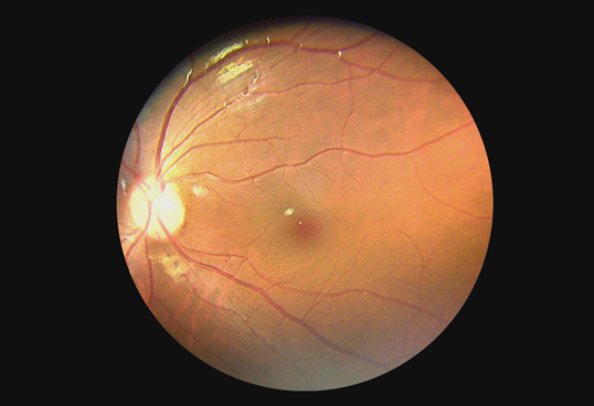
Image Gallery
Through the scope