Products

/Products/
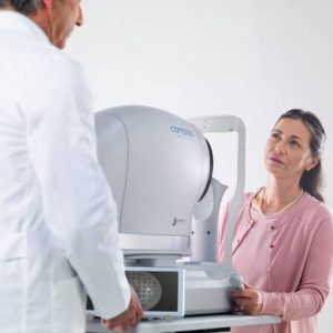
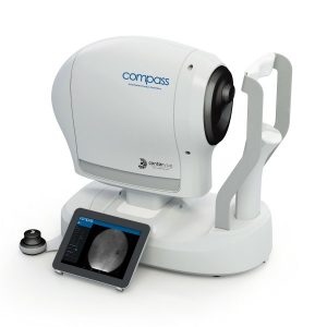
  • compass

    iCare COMPASS Automated Perimeter with active Retinal Tracking

    Key features

    • Standard automated perimetry
    • Active retinal tracking compensating for poor patient fixation in real-time
    • Auto-focus — no trial lens needed
    • Hygienic design
    • Illustrative fixation analysis; fixation area and plot
    • High-resolution confocal TrueColor imaging of the retina
    • No dilation of pupil needed, the patient can blink freely and the test can be suspended at any time without data loss
    • Ease of use & minimal operator training
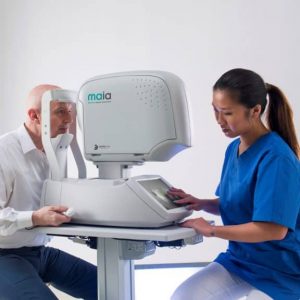
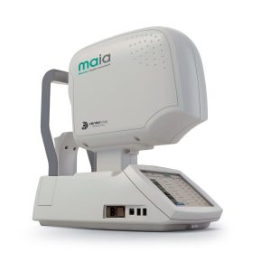
  • maia-modello

    iCare MAIA and S-MAIA confocal microperimeters

    Key features
    • 25 Hz eye-tracking with confocal SLO imaging
    • Pre-programmed and custom tests
    • Sensitivity and fixation indexes, illustrative sensitivity, and fixation maps
    • Informative examination and progression reports
    • 2.5 mm minimum pupil size
    • Patients with cataract (up to grade 3+) and media opacities can be examined
    • Auto-focus (from -15D to +10D)
    • Sensitive to functional changes due to macular pathologies or treatments even in early stages (background down to <0.0001 asb, threshold range 36 dB)
    • Simple to use, patients can be tested in less than 3 minutes per eye
    • S-MAIA includes also scotopic testing with cyan and red stimuli
     
  • iCare DRS

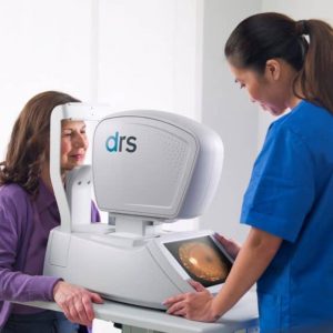
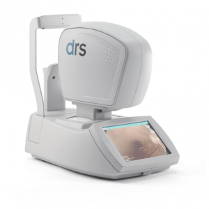
    iCare DRS automated digital retinography system

    Key features
    • Fully automated: patient autosensing, auto-alignment, auto-focus, auto flash adjustment, auto-capture
    • Short exam time
    • High quality color and Red-free images
    • Minimal training required
    • Compact and clean design
    • No additional PC required
    • Touch-screen operation
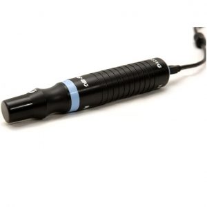
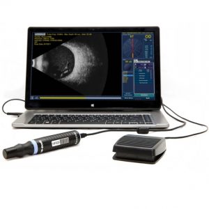
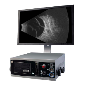
  • The B-Scan Plus’ and B-Scan Plus Vet's proprietary software combined with unique probe electronics make it the only portable High Definition B-Scan. While developing the B-Scan Plus software, Accutome visited many of the top facilities around the world. The results are a reliable and easy-to-use unit, which can quickly scan patients and transport information. Features:
    • 0.015 mm resolution is among the highest in the medical industry.
    • State-of-the-art probe design is sharper, more focused images due to the elimination of signal loss.
    • Contains 4 useful measuring tools along with corresponding A-Vectoring for diagnosing ocular pathologies.
    • The only unit with 2x full image zoom without distortion of real-time or captured scan.
    • Portable probe can plug into any laptop or PC.*
    • Unlimited 34 second film loops.
    • Share Information Easily with adaptable document transfer via EMR, email or printer.
    • Compact file storage is unsurpassed by the competition.
    • Protect your investment with easily upgradeable software.
    • User-friendly interface makes user manuals and quick start guides unnecessary.
    • The B-Scan Plus sofware is now DICOM compatable.
    * Certain Computer Specifications Required.
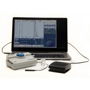
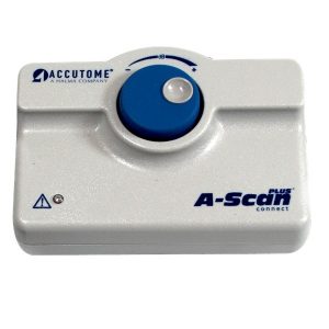
  • The A-Scan Plus Connect® is designed to meet the demands of today’s refractive cataract surgery.  It is the single most effective solution for measuring and calculating all of your patients and refining your surgical outcomes.  It has the ability to measure most patients and works great with dense cataracts or patients with fixation difficulties. The Connect links directly to a PC, laptop or tablet device making it the ultimate portable solution. Information can easily be uploaded to electronic medical record (EMR) systems. The improved user interface makes entering patient data, capturing the scan and calculating the measurement much faster.* Features:
    • Immersion and Contact Modes.
    • Industry Leading Resolution.
    • User-friendly interface.
    • Less Patient Chair Time – Faster measurement capture*.
    • Automatic Alignment Detection and Sclera Recognition eliminate marginally aligned scans.
    • Portable, Lightweight Design – Plug into any PC, laptop or tablet device.
    • Share information easily – adaptable document transfer via EMR, email or printer.
    • Modern Third and Fourth Generation Formulas including the Hoffer®Q, SRK/T, Holladay, Haigis as well as new post refractive formulas.
    • Optimized Lens Constants for superior surgical outcomes.
    • Unlimited Software Licenses.
    • Streamline your patient data - shares information with B-Scan Plus and UBM Plus.
    *When compared to original A-Scan Plus
  • The intelligence inside

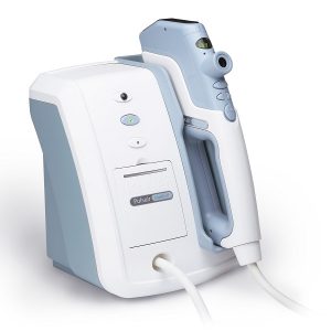
    The Keeler intelligent puff system giving you a quantum leap forwards in accuracy and ease of use. intelliPuff® embodies electronic and optical technology to deliver you the speed, accuracy and ease of use you and your patients deserve.

    Our softest puff ever

    Gentle is understating the new system - and if the patient has high pressures the puff will automatically increase for the next measurement. Kind, intelligent and simple. Technology working for you.

    Desktop or wall mount

    Save valuable space Pulsair intelliPuff® uses less than 50% of the space of conventional non contact tonometers and is the only model that can be wall mounted. Whether you wall mount or prefer to place it on your desk you win the 'space war' every time. No special instrument table is required - put it where you want it. Move it when you want to.  
  • ultra q reflexultra q reflex
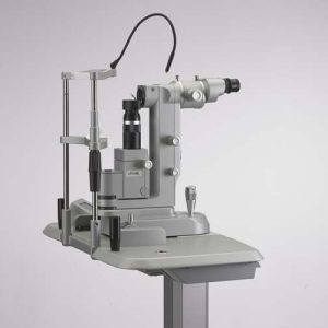
    ultra q reflexThe new Ultra Q Reflex™ is the only YAG laser designed to perform both anterior and posterior YAG treatments – and is ideally suited for the treatment of vitreous strands and opacities. 1. Capsulotomy with New Generation IOLs 2. Laser Vitreolysis for the Treatment of Floaters 3. Peripheral Iridotomy for Glaucoma

    Product Information

    1. Optimized Optics for Better Viewing 2. Reflex Technology 3. IOL-Friendly Photodisruption 4. Better Performance Over the Long-Term 5. Pinpoint Precision 6. Accurately Position Optical Breakdown 7. Designed to Maximize Your Workflow 8. Faster Treatment
  • Eyecubedellex Eyecubed

    eye-cubed

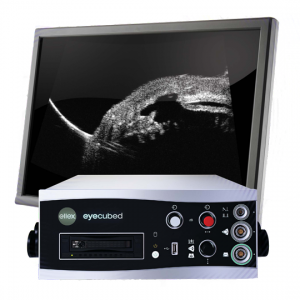
    Eye Cubed™ features real time imaging, advanced movie mode, real-time image enhancement, and a range of self-calibrating "best fit" probes.

    Customized Configuration to Best Meet Your Needs

    With customized configuration of A-Scan and B-Scan modes, Eye Cubed™ covers all of your diagnostic ultrasound needs for both the posterior and anterior segments. Pre-op or post-op, A-Scan or B-Scan, retina or anterior segment: whatever your focus, Eye Cubed™ shows you more, in more detail, than any other device of its kind.

    High Resolution Goes Ultra

    Eye Cubed’s 40 MHz UBM mode allows you to view anterior structures such as the cornea, iris, ciliary body, crystalline and intraocular lens more clearly than ever before. Whether determining the sulcus-to-sulcus measurement for accurate ICL sizing or the angle for potential angle closure and possible YAG laser iridotomy, Eye Cubed’s 40M Hz UBM mode is the gold-standard in high-resolution ultrasound.

    Essential Technology for Your Practice

    Even in an era of high-tech OCT scanning and digital imaging, ultrasound is the only means to obtain a crucial view of the posterior segment when there is a dense cataract or vitreous hemorrhage in the eye – making it one of the most fundamental diagnostic tools in ophthalmology. Detection of disorders like posterior vitreous detachment (PVD) in opaque ocular media is easily achieved with B-Scan ultrasound.

    Innovative Imaging

    Since acquiring Sacramento-based ophthalmic ultrasound pioneers Innovative Imaging Inc. in 2006, Ellex has been working hard to provide ever-increasing value to our Eye Cubed customers – and to develop our ultrasound technology platform. With the support of personalized, clinical ultrasound applications training by expert ecographers, Eye Cubed™ offers a total solution for your practice – and for your patients.

    Ophthalmic Edge

    Visit the comprehensive online diagnostic teaching tool created by Yale Fisher, MD, to access lectures, monographs and cases studies covering a range of diagnostic technologies, including ultrasound and OCT.
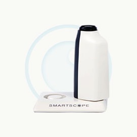
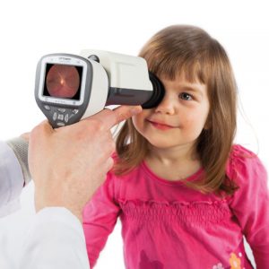
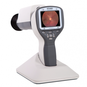
  • DIGITAL FUNDUS IMAGING Smartscope PRO camera with Smartscope FA module is a perfect combination of Slit Lamp and hand-held mode providing high quality Fluorescein Angiograms with a wide 40 degrees field of view. 9 internal fixation targets enable peripheral imaging (total FOV 70 * 52 degrees). Smartscope FA is easy to operate and offers fast image capture ability and a detailed view of the entire fluorescein dye circulation dynamics. With a special Slit Lamp Adapter Smartscope FA can be mounted to any Slit Lamp with patient head rest. Wi-Fi enables easy image transfer to PC, laptop, tablet or mobile devices. With Optomed Workstation software (sold separately) the images can be viewed, shared and stored locally and sent directly to a DICOM compatible system (PACS or other) of the hospital network.
  • optomed smartscope prooptomed smartscope pro
    DIGITAL FUNDUS IMAGING Optomed Smartscope® PRO is the next generation hand-held medical camera that provides high image quality which fulfills international ISO 10940 fundus camera standard requirements. Interchangeable modular optics make the camera even more versatile. The lightweight camera incorporates advanced technology in a portable design. Smartscope PRO camera provides accurate and silent autofocus which makes image capturing quick and easy. The camera is also equipped with Wi-Fi for advanced image transfer to any PC or mobile device. The Smartscope PRO comes with Patient Data Management software (sold separately) with full DICOM and PACS compliance to ensure seamless interoperability with hospital networks and practice management systems.
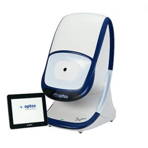
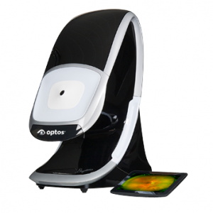
  • Our cutting edge retinal imaging technology is customized to fit the needs of your practice. The next generation of Optos technology, Daytona has been scaled to accommodate smaller office spaces while providing ultra-high resolution imaging, and adding ultra-widefield autofluorescence capabilities. Features:
    • Non-mydriatic ultra-high resolution images in under a second, through 2 mm pupils and many cataracts
    • Red and green lasers; each wavelength provides information for interpretation and diagnosis. Channels can be viewed separately:
      • Green (532 nm) “red-free” visualizes the sensory retina to the RPE
      • Red (635 nm) shows deeper structures of the retina (RPE to Choroid)
    • Ultra-widefield autofluorescence imaging with green laser light displays lipofuscin in the RPE
    • Images are available immediately and stored electronically for future comparison or telehealth applications
    • Innovative software tools enhance image evaluation
    • DICOM compatible
  • Optos-CaliforniaOptos-California
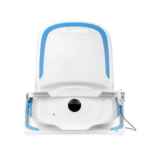
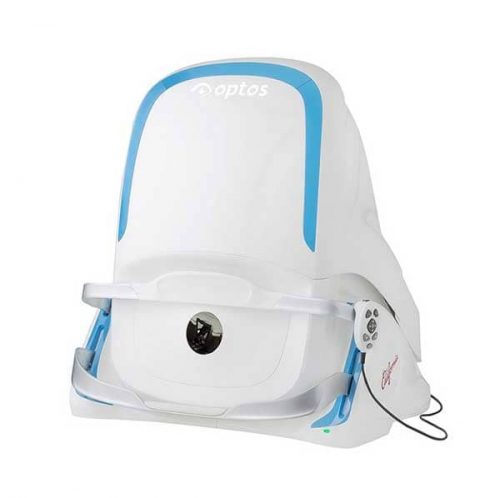
    With California, Optos has incorporated  new hardware and software technology enabling practitioners  to see more, discover more and effectively treat more ocular pathology thus promoting patient health.  We are committed to further strengthening our clinical evidence while demonstrating the importance of imaging the entire retina. We have created three versions of California to be customizable for use in multiple eyecare settings. California includes a new UWF optomap imaging modality; Indocyanine Green angiography (icg) while retaining:
    • Composite color
    • Red-free
    • Autofluorescence (af)
    • Fluorescein angiography (fa)
    Images are now presented in ProView which displays optomap in a consistent geometry that accurately represents anatomical features in the retina. Further, ProView enables automatic image registration for disease tracking over time, and inter-modality image comparison. New proprietary optical hardware optimizes and maintains resolution of the optomapimages throughout the scan of the retina resulting in more clarity in the far periphery. Image overlay enables comparison between composite color images and red-free, affa, or icg images.  Additionally, comparisons can be made between different images or different dates by scrolling through all stored images.