-
 The L-0189 5 STEPS MAGNIFICATION SLIT LAMP provides practitioners with the utmost satisfaction thanks to its excellent image clearness and fine design.
 -
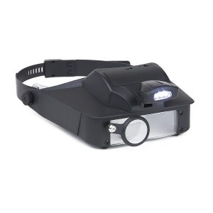 The LV-10 LumiVisor™ is a hands free, head-worn magnifier with 4 different powers; 2x, 3x, 5x and 6x . It comes with a fully adjustable one size fits all head strap. The LED light is fully adjustable so you can shine light wherever it is needed. The LumiiVisor™ magnifier is perfect for all hobbies, crafts, repairs and reading! The MagniVisor™ Deluxe is also ideal to be used as a low vision aid.
 -
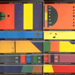 Basic Vertical Dr. Only Starter for any patient into fusion, anti-suppression and ranges of fusion. Set of 10 Cards For use with Bernell-O-Scope™ Item #: SMBV
 -
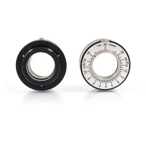 Fits into the standard 38 mm eyewell of the Bernell-O-Scope or any standard trial lens frame. Each prism yields 20 PD for a total of 40PD. Now you can set any amount of BI, BO, or vertical prism into the stereoscope to do training procedures. May be used for distance training by removing the cardholder. Item #: ARPHH20
 -
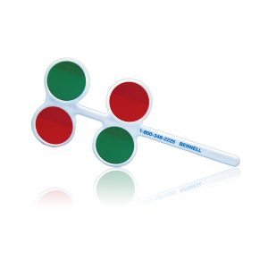 Red/Green Flippers Quality, four-well flipper bar features two alternating sets of red and green lenses. Reverses Base-In/Base-Out demand with each flip. Item #: BC1270RG
 -
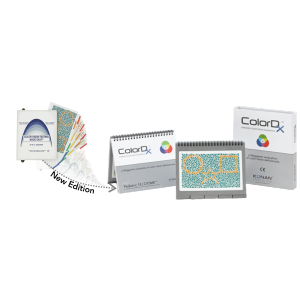 Same great test, but in a new and improved package! The ColorDx Pediatric 15 is the newest version of Color Vision Testing Made Easy or CVTME. As the Gold Standard pediatric assessment of color deficiencies or color blindness See The Multi-Ethnic Pediatric Eye Disease Study published by the AAO, CVTME™ is used in over 40 countries and has been validated for illiterates at the US Special Olympics. Featuring the proven Waggoner six confusion-color strategy, the pediatric test is used in a broad range of settings: pediatrics, eye physicians, school nurses, and many other disciplines of health care, in any language, world-wide. This easy to administer test features both hidden and non-hidden shapes that allow for positive reinforcement even while testing color deficient children by allowing successful answers on most targets. This success-reinforced strategy is also useful for assessment of malingering. Designed by color vision expert Dr. Terrace Waggoner, this new edition features enhanced digital colorimetery production methods, durable page and binding materials, and magnetic light-protective storage case and enhanced digital colorimetery production method and durable page materials and binding. Brilliantly simple to take, easy to score and just plain fun for children.
 -
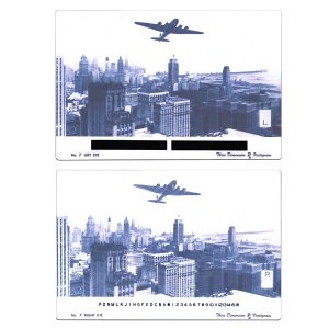
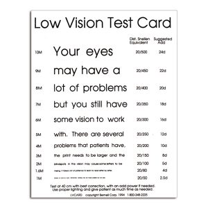 Features both upper and lower case letters to test near visual acuity in low vision patients. Appropriate starting high plus add power or magnifier power is indicated to the right of the line of copy the patient is able to read.
 -
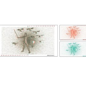
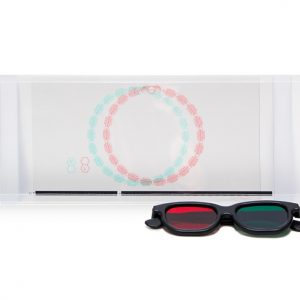
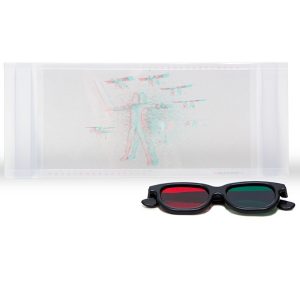 Slayer 3D Variable Tranaglyph. Replaces the popular Topper Vectogram. Size: 10.5" x 5.5" Material: Red/Green slides on transparent vinyl Usage: Can be used with Dual Polachrome Trainer or other slide holders.
 -
 8-1/4" x 5-1/2" Polarized vectograms ensure testing and training at actual distances. vectograms stabalize fusion and steropsis, eliminate suppression and develop simultaneous vision. Used in monocular, binocular, pursuit and saccadic training, vectograms help correct and prevent anomalies involving projection and hand-eye coordination. Ideal for use with Bernell's Dual Polachrome™ Trainer.
 -
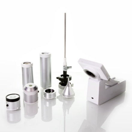
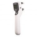
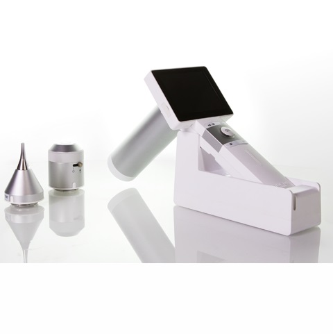 This innovative product “Digital Hand-held diagnostic set” is Class-II (ophthalmoscope)/Class-I (otoscope and dermoscope) Medical Device. It is expected to substitute for the traditional ophthalmoscope, otoscope and dermoscope by the use of the digital photographic solution. This medical device is provided to capture the digital photograph or video of eye-fundus, ear canal and tympanic membrane, epidermis and dermis of skin. Following the global trend of electronic medical records and telehealthcare networks, this people-oriented medical imaging product will be widely applied in doctors’ offices, clinics, skilled nursing facilities and healthcare station.
 -
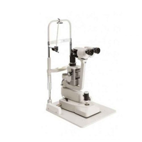
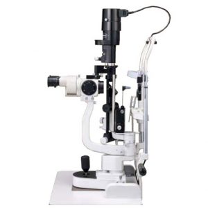
LED Slit Lamp RS Series
Slit lamp RS series have high quality optics with versatile functionalities and are easy to use. The series consists of three models: RS-5000, RS-500 and RS-300. RS-5000 is the high illumination tower model, while RS-500 is the low illumination compact model with 5 step magnifications. RS-300 is the primary model with three step magnifications yet with similar level of high performance and features. The optics of RS-5000/RS-500/RS-300 are bright and wide with an optimum convergence angle*(12 degrees). Drastic improvements were applied to the device in order to achieve comfortable and smooth operation by newly designed joystick and ergonomic base design that ensures solid movement to the various desired positions. The newly designed main and background illumination control dials are designed to be easily and intuitively used with one hand. From the wide range of LEDs, we have chosen the LED which is closest to the conventional halogen light colour temperature. *convergence angle is for RS-5000/500 only -
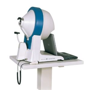
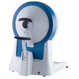 Thanks to its ergonomic design, user-friendly program navigation and ease of operation, the OCULUS Centerfield® 2 Perimeter has proven itself to be an invaluable instrument in the Occupational Health area. The unit performs static perimetry up to 70° eccentricity. It also meets the requirements of the German Ophthalmological Society’s (DOG) Road Traffic Commission for conducting visual field testing in accordance with the regulations for the issuance of driver’s licenses and the Guidelines G25 and G41 issued by the German Institute for Occupational Health and Safety.
Ergonomic Design
The self-contained design and light-shielded viewer allow examinations to be conducted in normally lit rooms. Its robustness and light weight make the OCULUS Centerfield® 2 Perimeter the ideal unit for portable use, which is often a necessity in the Occupational Health area.Reliable Results
The results of the perimetric measurements are summarized in a clearly structured printout. Areas of abnormality can be quickly recognized and re-examination of these areas independent of the test point grid gives the diagnostic analysis even greater reliability.Modern Connectivity
The OCULUS Centerfield® 2 Perimeter can be operated via the USB port of a notebook or PC. Together with the supplied device software, this modern solution allows you to fully utilize all of the benefits of today’s IT systems, especially the network connectivity. This guarantees both safe storage of examination data and quick access to that data when needed. -
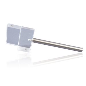
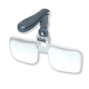 The VM-14 VisorMag™ from Carson Optical is a set of clip-on, flip-up magnifying glasses that attach to the brim of most caps and hats. Perfect for fly-tying and all fly-fishing needs. Complete with protective soft-pouch, its crystal clear acrylic Lenses give you a sharp, distortion-free view that allows you to see even the finest details with ease. These Magnifying glasses are 2.25x power equivalent to +5.00 diopter. Keep these clip-on Magnifier lenses in your tackle box so you’’ always have them at the ready.” Also available 1.75x power/+3.00 diopter: VM-10 2x power/+4.00 diopter: VM-12 US Patent D553,178
 -
 A good binocular screening device. Performed at a distance of 40 cm with a target of 20/30. Most normal patients can cycle between these prims 15/minute. Those with difficulty should be more thoroughly tested. 12D Base-Out and 3D Base-In. Prism is 1.5 inches square. Item #: G1110+
 -
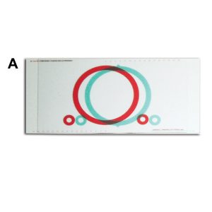
 New 3D Modeled Variable Tranaglyph. Comparable to the popular Quoit Vectogram.
 -
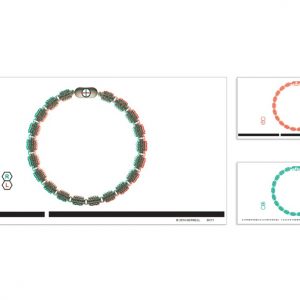
 Variable Tranaglyph™ Kit (500 Series) Variable prismatic red/green slides help build initial fusional reserves through peripheral and central fusion as well as stereopsis. Effective home training slides feature 5.5" x 10.5" red/green targets on transparent vinyl. Item #: BC500+
 -
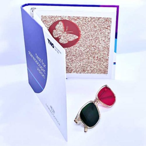 The TNO stereotest (19th edition) contains six test plates, presented in book form.
 -
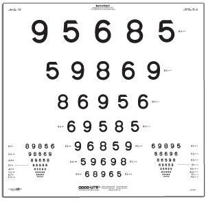 Adult LEA NUMBERS® chart for ETDRS Illuminated light boxes. Fits all existing ETDRS cabinets including both the ESV3000 and ESC2000. LEA NUMBERS® ETDRS chart contains optotypes in proportionally spaced (logMAR) lines; lines range from 20/200 to 20/8 (6/60 to 6/2.4) equivalent, 0.10 to 2.50. Fits in ESV3000 ETDRS illuminated cabinet.
 -
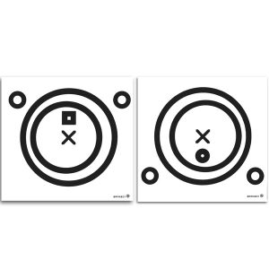
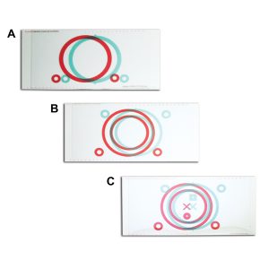
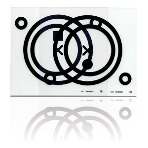 Free space fusion performed in a new, inexpensive way. Black target circles are printed on two 6" x 7" clear acetate cards. A unique, accurate method of developing increased fusional range without the need for costly lenses or filters.
 -
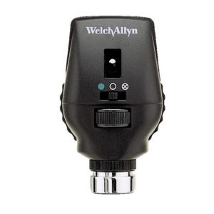 Patented Welch Allyn Coaxial Vision optics, combined with 68 lenses in single-diopter steps, for the precision you need to conduct a quality eye exam.
 -
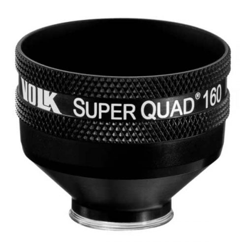 PART #VSQUAD160 Experience wide field, distortion-free visualization of the retina from the nerve head and macula, up to the ora serrata, designed for detection and treatment of retinal abnormalities like peripheral retinal tears, peripheral retinal detachments, giant retinal tears etc. The 30 mm lens surface offers a large, clear image of the retina for accurate and easy placement of the laser spot. The contact surface is designed carefully to provide optimum stability on the patient’s cornea while ensuring patient comfort.
- Wide-field distortion free viewing
- Ideal for detecting and treating mid to far-peripheral retinal abnormalities
- Large lens surface area providing a large working area
- Available in: Flanged contact, no Flange contact design
-
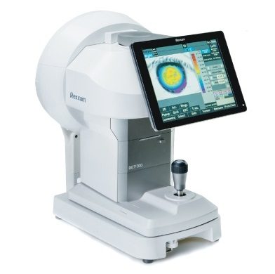
Multi functional all in one
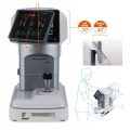
Auto Ref-Topographer RET-700 combines all the necessary features for anterior eye examination in just one device. RET-700 is not only a Topographer but Auto Refract Keratometer, Wavefront, Dry eye analysis (optional) and Meibomian glands analysis as well. -
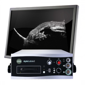
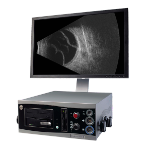
Eye Cubed™ features real time imaging, advanced movie mode, real-time image enhancement, and a range of self-calibrating "best fit" probes.
Customized Configuration to Best Meet Your Needs
With customized configuration of A-Scan and B-Scan modes, Eye Cubed™ covers all of your diagnostic ultrasound needs for both the posterior and anterior segments. Pre-op or post-op, A-Scan or B-Scan, retina or anterior segment: whatever your focus, Eye Cubed™ shows you more, in more detail, than any other device of its kind.High Resolution Goes Ultra
Eye Cubed’s 40 MHz UBM mode allows you to view anterior structures such as the cornea, iris, ciliary body, crystalline and intraocular lens more clearly than ever before. Whether determining the sulcus-to-sulcus measurement for accurate ICL sizing or the angle for potential angle closure and possible YAG laser iridotomy, Eye Cubed’s 40M Hz UBM mode is the gold-standard in high-resolution ultrasound.Essential Technology for Your Practice
Even in an era of high-tech OCT scanning and digital imaging, ultrasound is the only means to obtain a crucial view of the posterior segment when there is a dense cataract or vitreous hemorrhage in the eye – making it one of the most fundamental diagnostic tools in ophthalmology. Detection of disorders like posterior vitreous detachment (PVD) in opaque ocular media is easily achieved with B-Scan ultrasound.Innovative Imaging
Since acquiring Sacramento-based ophthalmic ultrasound pioneers Innovative Imaging Inc. in 2006, Ellex has been working hard to provide ever-increasing value to our Eye Cubed customers – and to develop our ultrasound technology platform. With the support of personalized, clinical ultrasound applications training by expert ecographers, Eye Cubed™ offers a total solution for your practice – and for your patients.Ophthalmic Edge
Visit the comprehensive online diagnostic teaching tool created by Yale Fisher, MD, to access lectures, monographs and cases studies covering a range of diagnostic technologies, including ultrasound and OCT. -
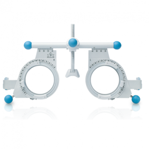
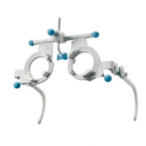 Everyone who has anything to do with the prescription of eyeglasses has heard of OCULUS Universal Trial Frames. Function
- Space for a total of 10 trial lenses of 38 mm diameter
- PD range from 46-80 mm
- Adjustable temple tilt and length
- Smooth axis adjustment
- CVD scale
+65 6514 0848|info@ophthalmic.com.sg