Typology: Tear Film
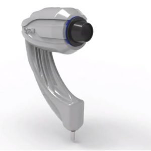
Acquisition Mode: Multi shot, tube,
ISO Management: Variable
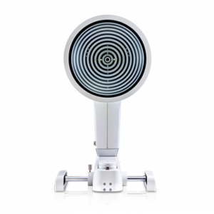
Grids: Placido disc, NIBUT grid
Filters: Yello Filter
Lighting: White LED – Blue LED
Image Resolution: 8-12 mp
Focus: Autofocus, Manual focus
Magnification: 4x to 8x


Typology: Tear Film
Acquisition Mode: Multi shot, tube,
ISO Management: Variable
Grids: Placido disc, NIBUT grid
Filters: Yello Filter
Lighting: White LED – Blue LED
Image Resolution: 8-12 mp
Focus: Autofocus, Manual focus
Magnification: 4x to 8x
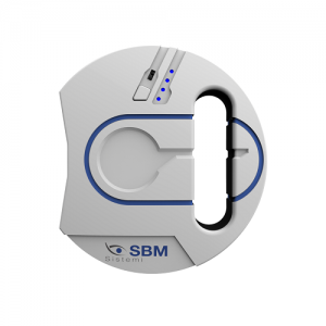
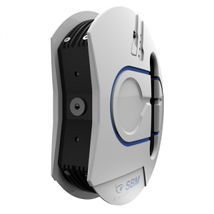
ICP Dry Eye analysis
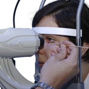
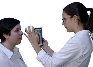
I.C.P Tearscope the new instrument of individual analysis of lacrimal that allow with a quick detailed structural research of the tear composition.
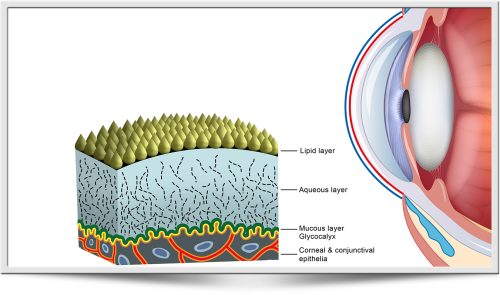
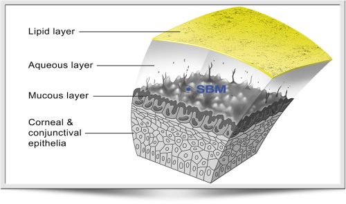
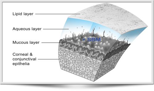
Possibility of researsch on the single layers:
Thanks to ICP Tearscope is possible to identify the type of DED(Drye Eye Desease) and determine which deficient layers can be treated with a specific treatment.
Thanks to ICP OSA is possible to identify the type of DED(Drye Eye Desease) and determine which deficient layers can be treated with a specific treatment.
Mucin layer and water layer analysis
The layer watery is evaluated on the meniscus tear categorizing in different categories and possible issues related this. The measurement in mm allows without invasiveness the direct evaluation of the quantity.
MGD Analysis meibomian gland desease
Easily and efficiently integrates complex examination, such as meibography into the ophthalmological and optometric practices.
Dry Eye is most commonly caused by the Meibomian Gland Dysfunction (MGD). The Meibo-Scan shows the morphological changes in the glandural tissue.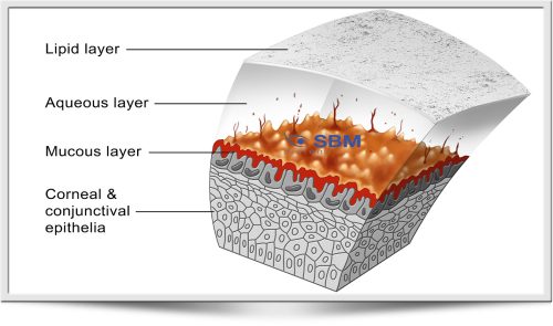
System analysis of the images obtained through a sisnsitive infrared camera (NIR) in order to locate in a guided way:
| Tipology: | Tear film |
| Image resolution: | From 8 to 12 mp |
| Acquisition mode: | Multi shot, tube |
| Focus: | Autofocus, manual focus |
| ISO management: | Variable |
| Magnification: | 4x to 8x magnification with change via software |
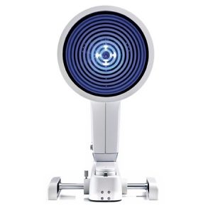
| Grids: | Placido disc, NIBUT grid |
| Filters: | Yellow filter |
| Lighting: | White led – Blue led |
I.C.P. Tearscope the new instru ment of individual analysis of lacrimal layers that allow with a quick detailed structural research of the tear composition.
Possibility of researsch on the single layers:
• Lipid
• Aqueous
• Mucin
Thanks to ICP Tearscope is possible to identify the type of DED(Dry Eye Desease) and determine which deficient layers can be treated with a specific treatment.
 With white LEDs lighting displays in vivo the phenomenon of interference fringes possible to assess the thickness of the lipid component of the tear and run the NiBUT.
With blue LEDs lighting (with the fluorescein) creates a large area and allows you to perform the BUT and look fluorescein of large diameter scleral and mini scleral contact lenses type.
 Through the use of GRADING SCALE dedicated to each value obtained from the exams, the interpretation of the obtained data results easy and immediate making the iPad a real platform dedicated to the analysis of dry eye with detailed temporal graphics that allow to demonstrate in simple steps the need of the treatments and then the effective functioning of these!
The layer watery is evaluated on the meniscus tear categorizing in different categories and possible issues related this. The measurement in mm allows without invasiveness the direct evaluation of the quantity.ugh the use of dedicated filters in real time after the acquisition.
 The system is provided with a kit of useful grids to perform various screening, all filters are already present in the system software and includes tests to evaluate and diagnose dry eye problems and can recommend artificial tears.
• Measurement of BLACK LINE (MLMI)
• Evaluation of the integrity of cornea and ascertaining the presence of corneal scars and bruises. The product is already ready for the connection to Digital Imaging and Communications in Medicine (DICOM)
Supplied accessories
• Blue and white Led
• A thick grid to observe the quality of the tear film and measure the N.I.B.U.T.
• A fine grid to evaluate the quality and the structure of tear
• A Placido’s disc to highlight possible distortions or corneal irregularities
• A yellow and cobalt blue filter via software for applicative evaluation of rigid contact lenses.
 Through a quick and easy acquisition of a series of 3 blinks, ICP Tearscope allows to obtain the thickness of the single Lipid Layer of the tear film classifying it in 7 different categories in a quick and precise way the secretion of the lipids by the Meibomian Glands.
Presence of grading scale and comparison in the time for detailed and precise follow up.
For a detailed analysis of the mucin layer, ICP Tearscope evaluates in both modes the break up time of the lipid layer and, so, the stability through the classic TBUT with possibilities of use of fluorescein in blue light the non-invasive and quick N.I.B.U.T.
 The tear film is the thin layer of liquid (about 8 μ, its thickness is variable on basis the considered portion and it results at maximum at cornea level) composed 98% of water and for the remaining 2% by protein and lipids, that is continuously and uniformly distributed on the ocular surface of the closing of the eyelids and that performs irreplaceable functions for our sight.
In fact it is able to improve the optic quality of the image regularizing the corneal surface (it has an index of refraction of 1,33, very close to that of the cornea); it allows an adequate lubrication reducing the friction of the eyelids, it allows the transport and the diffusion of molecules (oxygen, carbon dioxide, ions, mucins, lipids with a slightly alkaline pH 7,3/7,8), vital elements for the survival of the epithelia and of the cornea, it has strong antibacterial activity thanks to the presence of some enzymes and it guarantees the parts and keeps the ocular surface clean removing impurities from the environment, the waste of metabolism and exfoliated cells.