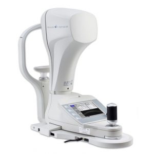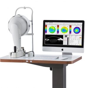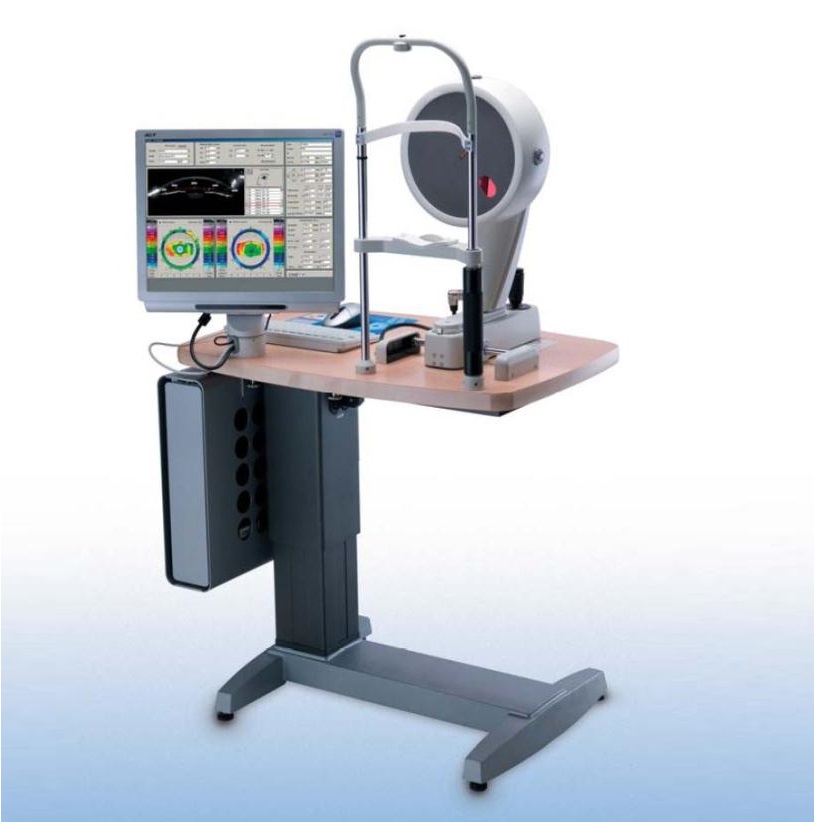


First introduced in 1999, the OCULUS Pentacam® is commercially available since 2003. It is the first automatically rotating Scheimpflug camera. During the rotating scan that takes max. 2 seconds, up to 50 Scheimpflug images of the anterior eye segment are captured. The examination is released automatically and is user independent. During the scanning process, the patient’s eye motions are captured using a second camera and compensated mathematically. Ray tracing is used to compensate for optical distortions. This combination is the basis for calculating solid data for further evaluation. More than 100 published studies and papers prove the efficiency of this concept. The OCULUS Pentacam® measures the cornea from limbus to limbus. It supplies topographic data on elevation and curvature of the entire anterior and posterior corneal surface. The corneal thickness (pachymetry) is measured and presented graphically over its entire surface. A topography based keratoconus detection and quantification are performed. The anterior chamber depth, chamber volume (size) and the chamber angles are calculated and presented for the Glaucoma screening. The illumination of the eye using blue LED light makes corneal and lens opacities (cataract) visible. For patients information, the anterior chamber can be visualized and displayed with the virtual tomography model. After the examination, OCULUS Pentacam® provides an indice report that summarizes the abnormalities found during the scan. This report is based on clinical published studies and articles that define abnormalities.
The OCULUS Pentacam® can be customized with two software packages and several software modules to fit your exact needs.
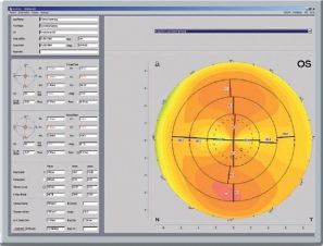 The rotating measurement principle guarantees the high resolution of the measuring points in the central cornea. Topographic analysis of the anterior and posterior corneal surfaces is based on the measured real height data. These provide the basis for:
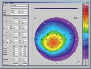 Pachymetry maps:
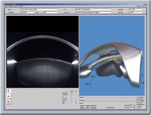 Applications:
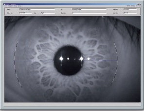 Applications:
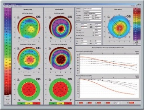
 Applications: