-
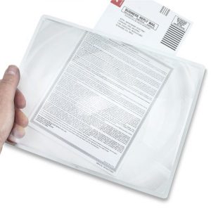
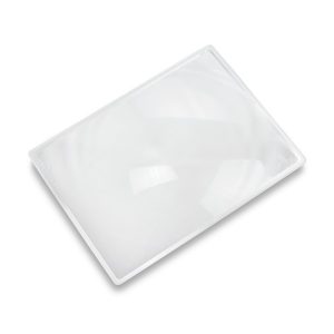 The DM-21 Page Magnifier from Carson Optical is a deluxe rigid frame full page magnifier. It’s 2x power acrylic fresnel lens is sturdy and shatterproof. The Page Magnifier is perfect for reading large areas of small print such as maps, newspapers, directories, recipes and more. Simply hold the Page Magnifier 4 inches above the reading material for a clear view. The Page Magnifier is so thin you can store it in your reading material after use.
 -
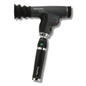 Our revolutionary PanOptic Ophthalmoscope addresses the fundamental challenge in ophthalmoscopy—to get a good view of the fundus in order to make a sufficient assessment. Patented Axial PointSource™ Optics make it easy to enter undilated pupils, offering a 25º field of view, resulting in a view of the fundus that's 5X greater than you see with a standard ophthalmoscope in an undilated eye. Direct viewing of the fundus through the PanOptic provides better images of the retinal changes caused by hypertension, diabetic retinopathy, glaucoma, and papilledema to enable clinicians to make these diagnoses earlier.
 -
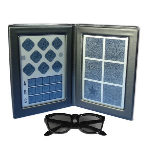 Paul Harris Randot Test (Special Edition) This test is recommended in lectures by Baltimore Academy for Behavioral Optometry. Special run by Stereo Optical exclusively for Bernell. Test down to 20 sec. Includes: Polarized Goggle
 -
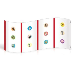
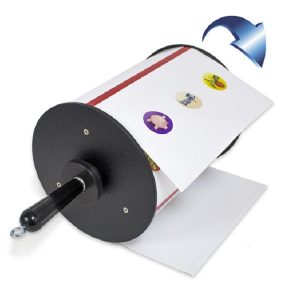 Sized to fit over our Adult Optokinetic Drum (DAL300) this vinyl sheet is used to test for the optokinetic reflex. The banner has child interest targets similar to our DAL301. Can be rolled up for easy storage. Clear end pieces hold banner from curling. Washable plastic material similar to window shade. Velcro on backside of the banner allows for easy attachment around the Adult Optokinetic Drum (DAL300). Item #: OKNADP1
 -
 Vectographic Projector Slide Pediatric Slide This pediatric projector slide test depth perception and binocularity under distance conditions. The top portion of each slide is a standard non-vectographic projector slide. The bottom portion is a vetographic slide that contains suppression control targets, a distance stereo acuity test, fixation disparity and binocular balance. Slide fits most AO and Marco projectors, and there are adaptable to a 10' or 20' lane. REQUIRES POLARIZING SCREEN.
 -
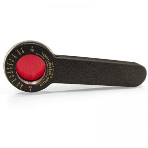 A Hand Held instrument to Measure a Maddox Phoria. This tests for anisophorias, vertical and horizontal, in all positions of gaze in real space instead of behind of phropter. Scale from 0 to 10 PD 25mm Item #: BC1211
 -
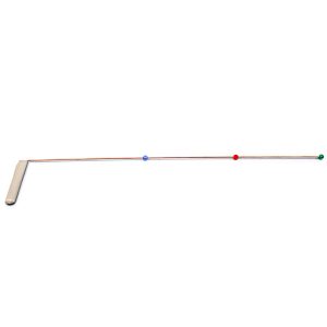 Copper Rod Ideal home training device for non-strabismics, esotropes with near centration point, and patients with other binocular vision problems. Three movable plastic beads on hand-held copper rod allows extensions of physiological diplopia techniques from 2" to 20'. Item #: BC1091
 -

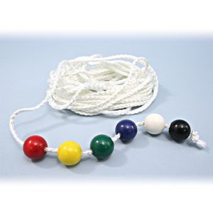 Physiological-Diplopia Cord™ (Similar to Brock String) Inexpensive home use instrument for near training as well as to extend phy-dip training from near into distance. Utilizes physical diplopia for training suppression, binocularity and spatial localization. Heavy cord with colored beads. Sold by the dozen in 10' or 20' lengths. Item #: BC109+
 -
 Plano Stick Prism Item #: ASPSPL
 -
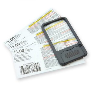
 The PM-33 Pocket Magnifier™ from Carson Optical is a multi-power LED lighted Magnifier. This HandHeld Pocket Magnifier™ has three magnifying powers: 6x, 5x and 2.5x. It features a crystal-clear acrylic lens. It is an ideal low vision aide. The Pocket Magnifier™ is also perfect for reading fine print. It is so compact that it can easily fit in a pocket or purse.
 -
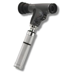
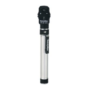 Patented Welch Allyn Coaxial Vision optics, combined with 68 lenses in single-diopter steps, for the precision you need to conduct a quality eye exam.
 -
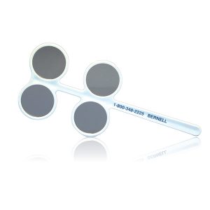 Four-well flipper bar features two sets of lenses to alternate demand. Used with Tranaglyphs or Vectograms to reverse BI/BO demands. Item #: BC1270POL
+65 6514 0848|info@ophthalmic.com.sg