- Avoid unnecessary mydriatic medications and side effects.
- Improve quality and efficiency of making the rounds and even reach those in remote areas.
- Minimize paperwork with images or videos taken, stored, and processed digitally.
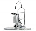
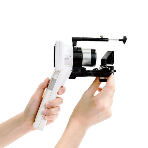
- Acquire additional stability with a special adapter attached to slit lamp.
-
-
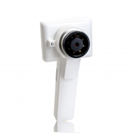
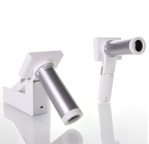
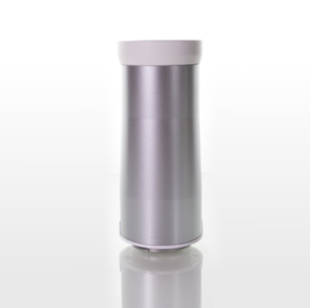
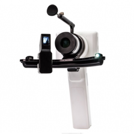
 Optomed Smartscope® PRO is the next generation hand-held medical camera that provides high image quality which fulfills international ISO 10940 fundus camera standard requirements. Interchangeable modular optics make the camera even more versatile. The lightweight camera incorporates advanced technology in a portable design.
Smartscope PRO camera provides accurate and silent autofocus which makes image capturing quick and easy. The camera is also equipped with Wi-Fi for advanced image transfer to any PC or mobile device.
The Smartscope PRO comes with Patient Data Management software (sold separately) with full DICOM and PACS compliance to ensure seamless interoperability with hospital networks and practice management systems.
Optomed Smartscope® PRO is the next generation hand-held medical camera that provides high image quality which fulfills international ISO 10940 fundus camera standard requirements. Interchangeable modular optics make the camera even more versatile. The lightweight camera incorporates advanced technology in a portable design.
Smartscope PRO camera provides accurate and silent autofocus which makes image capturing quick and easy. The camera is also equipped with Wi-Fi for advanced image transfer to any PC or mobile device.
The Smartscope PRO comes with Patient Data Management software (sold separately) with full DICOM and PACS compliance to ensure seamless interoperability with hospital networks and practice management systems. -
-
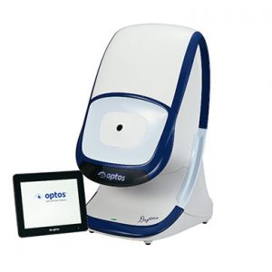
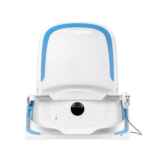
 Our cutting edge retinal imaging technology is customized to fit the needs of your practice. The next generation of Optos technology, Daytona has been scaled to accommodate smaller office spaces while providing ultra-high resolution imaging, and adding ultra-widefield autofluorescence capabilities. Features:
- Non-mydriatic ultra-high resolution images in under a second, through 2 mm pupils and many cataracts
- Red and green lasers; each wavelength provides information for interpretation and diagnosis. Channels can be viewed separately:
- Green (532 nm) “red-free” visualizes the sensory retina to the RPE
- Red (635 nm) shows deeper structures of the retina (RPE to Choroid)
- Ultra-widefield autofluorescence imaging with green laser light displays lipofuscin in the RPE
- Images are available immediately and stored electronically for future comparison or telehealth applications
- Innovative software tools enhance image evaluation
- DICOM compatible
-
-
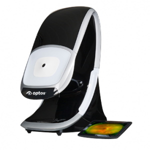
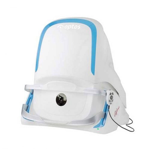 With California, Optos has incorporated new hardware and software technology enabling practitioners to see more, discover more and effectively treat more ocular pathology thus promoting patient health. We are committed to further strengthening our clinical evidence while demonstrating the importance of imaging the entire retina. We have created three versions of California to be customizable for use in multiple eyecare settings. California includes a new UWF optomap imaging modality; Indocyanine Green angiography (icg) while retaining:
- Composite color
- Red-free
- Autofluorescence (af)
- Fluorescein angiography (fa)
-
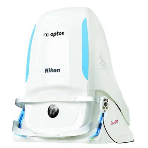 Silverstone, the most powerful tool yet for examining the retina, is the only ultra-widefield imaging device with integrated swept source OCT. Silverstone produces a 200° single-capture retinal image of unrivaled clarity in less than ½ second and enables optomap guided OCT scanning across the retina and into the far periphery.
-
 Portable Non-mydriatic eye fundus examination
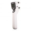
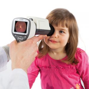
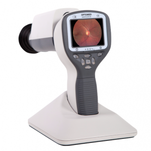
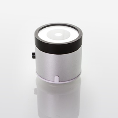
Optomed Smartscope M5 is a digital medical camera that provides general, ophthalmoscopic, otoscopic and dermatoscopic imaging with one hand-held device. This multipurpose digital imaging device weights only 400g and powered with a battery it gives you the freedom to move around and take the device with you to any location.
Digital still and video images created with Optomed Smartscope M5 allow making accurate first diagnosis and planning consistent follow-up treatment. Optomed Smartscope M5 is easily adopted into daily examination routines and connectivity to any patient database system enables fluent image data sharing e.g. for consultation purposes. The device comes with a 2GB SD memory card and pictures can be easily transferred to your PC by placing the device to its cradle which connects to the PC via USB connection.
The 5 megapixel sensor and up to 2560 x 1920 pixels resolution guarantee the quality of your pictures. Each of our 4 attachable optics modules has its own light source and adjustable illumination levels. Optics are fast and easy to attach and detach with bayonet connectors.
The advances in M5 compared to M3-2 are new sensor enabling improved picture quality, higher battery capacity and keyboard lights that make using the device in a dark room easier.
Optomed Smartscope M5 offers non-mydriatic fundus imaging without the need for dilation drops with EY2 and EY3 optics modules.
Portable Non-mydriatic eye fundus examination
Optomed Smartscope M5 is a digital medical camera that provides general, ophthalmoscopic, otoscopic and dermatoscopic imaging with one hand-held device. This multipurpose digital imaging device weights only 400g and powered with a battery it gives you the freedom to move around and take the device with you to any location.
Digital still and video images created with Optomed Smartscope M5 allow making accurate first diagnosis and planning consistent follow-up treatment. Optomed Smartscope M5 is easily adopted into daily examination routines and connectivity to any patient database system enables fluent image data sharing e.g. for consultation purposes. The device comes with a 2GB SD memory card and pictures can be easily transferred to your PC by placing the device to its cradle which connects to the PC via USB connection.
The 5 megapixel sensor and up to 2560 x 1920 pixels resolution guarantee the quality of your pictures. Each of our 4 attachable optics modules has its own light source and adjustable illumination levels. Optics are fast and easy to attach and detach with bayonet connectors.
The advances in M5 compared to M3-2 are new sensor enabling improved picture quality, higher battery capacity and keyboard lights that make using the device in a dark room easier.
Optomed Smartscope M5 offers non-mydriatic fundus imaging without the need for dilation drops with EY2 and EY3 optics modules. -
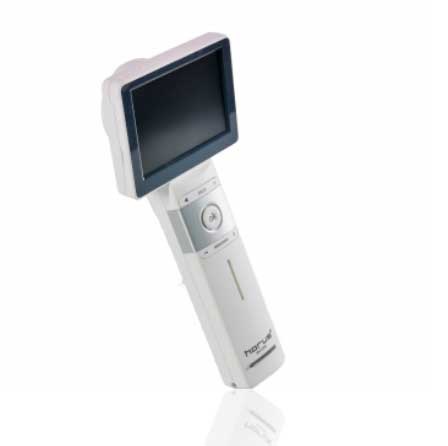
- Provide high definition clinical image
- Portable and hand held application in disease screen
- Friendly user interface with touch screen and Auto focus
- Multi functional diagnosis in ophthalmology , ENT . Dermatology and general practice.
- Widely application in clinic room , private office ,hospital, Tele- medicine , and mobile health.
-
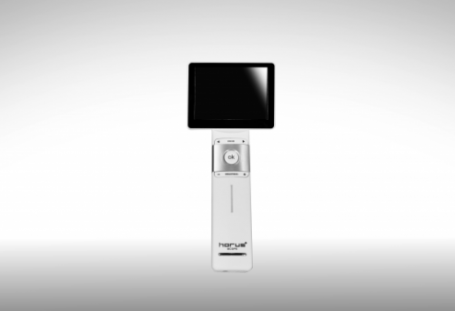
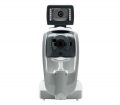
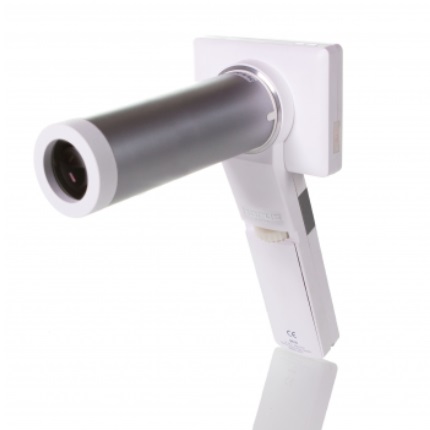
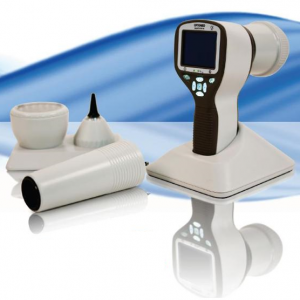
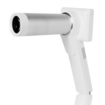 Horus DEC200 Non-Mydriatic Digital Handheld Fundus Camera offers high image quality with ISO 10940 fulfillment. 5MP (2592*1944 pixels) and 45 degree FOV of fundus image are captured to provide more details. 7 internal fixation targets for macula center, disk center and peripheral image in DEC 200 optical modules. Horus DEC200 provides both auto-focus and power-focus function to facilitate image capturing. Touch LCD Screen and Wi-Fi compatibility are also equipped . With a special slit lamp jig, Hours DEC 200 can be mounted with slit lamp for desktop application.
 -
-