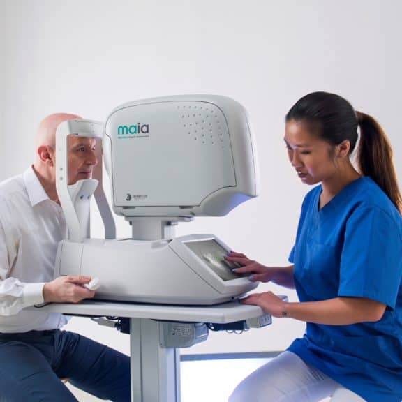
iCare MAIA and S-MAIA Microperimeters with Confocal Scanning Laser Ophthalmoscopy (SLO)
iCare MAIA (Macular Integrity Assessment)and S-MAIA offer the best in confocal microperimetry to combine visual field tests, fixation loss correction by a real-time retinal tracker and non-mydriatic confocal SLO fundus imaging, all in one exam. iCare MAIA and S-MAIA detect and monitor functional changes of the retina with great reliability.
iCare MAIA and S-MAIA perform different types of pre-programmed as well as customizable microperimetry tests and follow-up to monitor functional progression. The retinal tracker allows for accurate, real-time, calculation and compensation for eye movements. iCare MAIA and S-MAIA are patient friendly, automated, easy to use and non-mydriatic.
Microperimetry with an enhanced functional and structural correlation 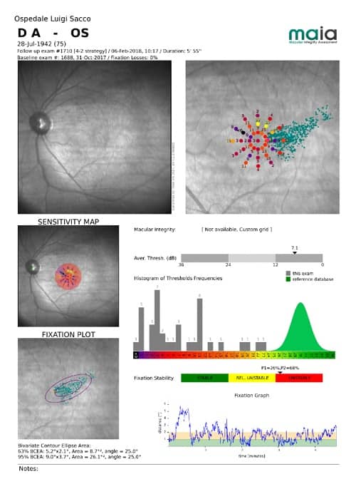
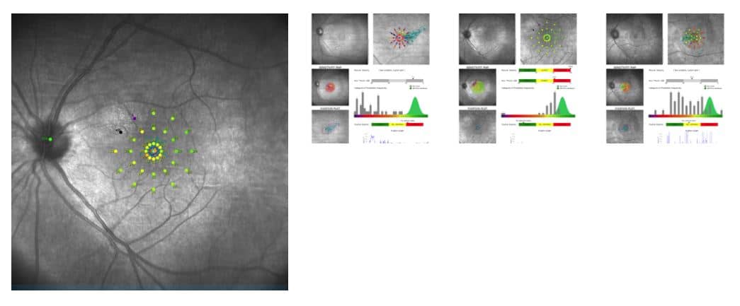
Microperimetry is a technology that allows concurrent analysis of structural and functional aspects of the retina. It combines retinal sensitivity mapping and fixation analysis with the fundus image in one exam as a powerful tool to detect, describe and follow-up pathologies affecting the macular area. iCare MAIA performs different types of microperimetry tests with supra and full-threshold strategies, and follow-up tests to monitor functional progression. Each exam provides a measure of retinal sensitivity and fixation analysis (stability and position of the Preferred Retinal Locus).
Fixation analysis for a better diagnosis and vision
iCare MAIA provides accurate and objective information regarding retinal location and stability of a patient’s fixation. Parameters are assessed by tracking eye movements 25 times per second and by plotting the resulting distribution over the SLO image. iCare MAIA can project multiple fixation targets at selectable locations to help patients visualize the goal during the sensitivity test. The fixation test can also be done alone and takes only seconds. Fixation test can also be conducted to train the patient to use a new optimal Preferred Retinal Locus.
iCare MAIA offers the best in confocal microperimetry
iCare MAIA performs different types of pre-programmed as well as customizable microperimetry tests and follow-up to monitor functional progression. The retinal tracker allows for accurate, real-time, calculation and compensation of eye movements.
Accurate and detailed diagnosis of macular diseases
iCare MAIA is used as a diagnostic device to aid in the detection and management of diseases affecting the macula, including, but not limited to, macular degenerations. Thanks to its combined structure-function analysis, microperimetry represents an essential tool for deriving the correct diagnostic decision in a variety of retinal diseases, monitoring the progression of retinal pathologies, assessing macular function prior to cataract surgery, examining patients with unexplained vision loss and more.
iCare S-MAIA enables scotopic testing
S-MAIA is the product version that can perform also scotopic tests for finding and explaining relation between the diseases and sensitivities of dark-adapted photoreceptor cells. In the scotopic mode the color of the projected stimuli can be chosen between cyan and red. The cyan scotopic test aims to stimulate the rod photo-receptors and to measure their sensitivity. The red scotopic test aims to measure the response of the red cone photo-receptors, without any interference due to the response of the rods.
Great ergonomics for a better experience
iCare MAIA and S-MAIA offer significant ergonomic advantages in terms of easy interaction between the operator, the patient, and the device. Thanks to the SLO, they operate with a minimum pupil diameter of 2.5 mm, not requiring the use of dilating drops, and it automatically corrects refraction errors from -15D to +10D. The devices have an automatic OD/OS recognition and an alarm that advises the operator when the patient is not properly following the test. The system is entirely operated via the integrated 10.4” touch-screen with 1024×768 resolution. The integrated PC offers dual USB and Ethernet interfaces.