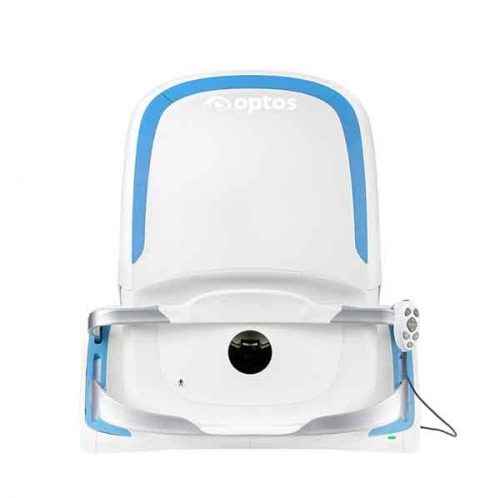
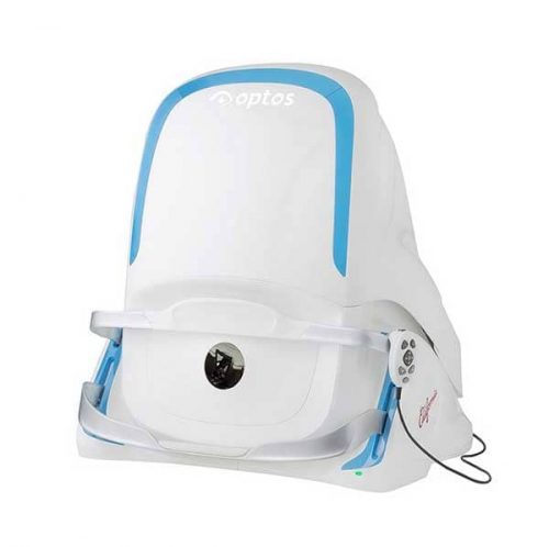
Imaging Tool

//Imaging Tool
  • Horus Digital Eye Anterior Lens_DEA 200Horus Digital Eye Anterior Lens_DEA 200
  • Horus Eye Surface Camera_DGC 200
  • Horus Portable Chin Rest_CR 100
  • iCare_DRSplus

    iCare DRSplus TrueColor confocal fundus imaging system

    Key features
    • TrueColor Confocal Technology
    • Multiple imaging modalities including red-free, external eye and stereo view imaging
    • 2.5 mm minimum pupil size
    • Fast, easy and fully automated operations
    • Mosaic function which creates retinal panoramic views up to 80°
    • Remote Viewer that allows for reviewing from devices on the same local area network
    • Remote Exam feature enables executing an exam from a distance
  • Horus Digital Diagnostic Set_DSC 100Horus Digital Diagnostic Set_DSC 100
    This innovative product “Digital Hand-held diagnostic set” is Class-II (ophthalmoscope)/Class-I (otoscope and dermoscope) Medical Device. It is expected to substitute for the traditional ophthalmoscope, otoscope and dermoscope by the use of the digital photographic solution. This medical device is provided to capture the digital photograph or video of eye-fundus, ear canal and tympanic membrane, epidermis and dermis of skin. Following the global trend of electronic medical records and telehealthcare networks, this people-oriented medical imaging product will be widely applied in doctors’ offices, clinics, skilled nursing facilities and healthcare station.
  • DIGITAL FUNDUS IMAGING Smartscope PRO camera with Smartscope FA module is a perfect combination of Slit Lamp and hand-held mode providing high quality Fluorescein Angiograms with a wide 40 degrees field of view. 9 internal fixation targets enable peripheral imaging (total FOV 70 * 52 degrees). Smartscope FA is easy to operate and offers fast image capture ability and a detailed view of the entire fluorescein dye circulation dynamics. With a special Slit Lamp Adapter Smartscope FA can be mounted to any Slit Lamp with patient head rest. Wi-Fi enables easy image transfer to PC, laptop, tablet or mobile devices. With Optomed Workstation software (sold separately) the images can be viewed, shared and stored locally and sent directly to a DICOM compatible system (PACS or other) of the hospital network.
  • Horus Digital Fundus Camera DEC 100Horus Digital Fundus Camera DEC 100
    • Avoid unnecessary mydriatic medications and side effects.
    • Improve quality and efficiency of making the rounds and even reach those in remote areas.
    • Minimize paperwork with images or videos taken, stored, and processed digitally.
    • Acquire additional stability with a special adapter attached to slit lamp.
  • optomed smartscope prooptomed smartscope pro
    DIGITAL FUNDUS IMAGING Optomed Smartscope® PRO is the next generation hand-held medical camera that provides high image quality which fulfills international ISO 10940 fundus camera standard requirements. Interchangeable modular optics make the camera even more versatile. The lightweight camera incorporates advanced technology in a portable design. Smartscope PRO camera provides accurate and silent autofocus which makes image capturing quick and easy. The camera is also equipped with Wi-Fi for advanced image transfer to any PC or mobile device. The Smartscope PRO comes with Patient Data Management software (sold separately) with full DICOM and PACS compliance to ensure seamless interoperability with hospital networks and practice management systems.
  • EEC-100
  • Our cutting edge retinal imaging technology is customized to fit the needs of your practice. The next generation of Optos technology, Daytona has been scaled to accommodate smaller office spaces while providing ultra-high resolution imaging, and adding ultra-widefield autofluorescence capabilities. Features:
    • Non-mydriatic ultra-high resolution images in under a second, through 2 mm pupils and many cataracts
    • Red and green lasers; each wavelength provides information for interpretation and diagnosis. Channels can be viewed separately:
      • Green (532 nm) “red-free” visualizes the sensory retina to the RPE
      • Red (635 nm) shows deeper structures of the retina (RPE to Choroid)
    • Ultra-widefield autofluorescence imaging with green laser light displays lipofuscin in the RPE
    • Images are available immediately and stored electronically for future comparison or telehealth applications
    • Innovative software tools enhance image evaluation
    • DICOM compatible
  • DEA-100 (Gonioscope)
  • Optos-CaliforniaOptos-California
    With 
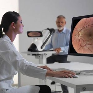
California, Optos has incorporated  new hardware and software technology enabling practitioners  to see more, discover more and effectively treat more ocular pathology thus promoting patient health.  We are committed to further strengthening our clinical evidence while demonstrating the importance of imaging the entire retina. 
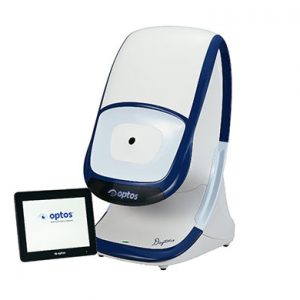
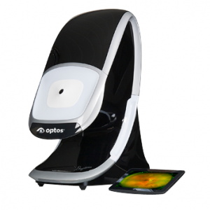
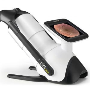
We have created three versions of California to be customizable for use in multiple eyecare settings. California includes a new UWF optomap imaging modality; Indocyanine Green angiography (icg) while retaining:
    • Composite color
    • Red-free
    • Autofluorescence (af)
    • Fluorescein angiography (fa)
    Images are now presented in ProView which displays optomap in a consistent geometry that accurately represents anatomical features in the retina. Further, ProView enables automatic image registration for disease tracking over time, and inter-modality image comparison. New proprietary optical hardware optimizes and maintains resolution of the optomapimages throughout the scan of the retina resulting in more clarity in the far periphery. Image overlay enables comparison between composite color images and red-free, affa, or icg images.  Additionally, comparisons can be made between different images or different dates by scrolling through all stored images.
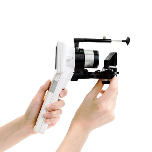
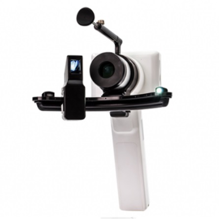
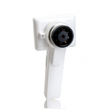
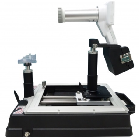
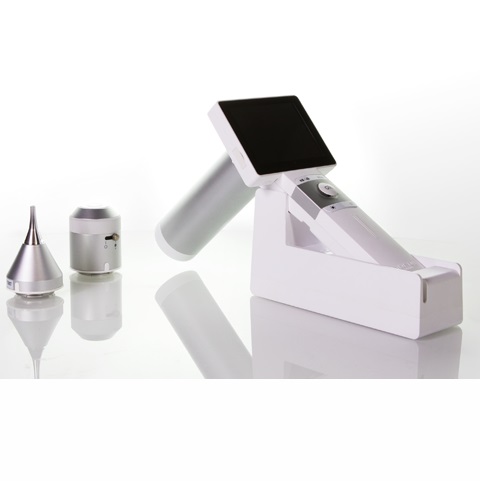
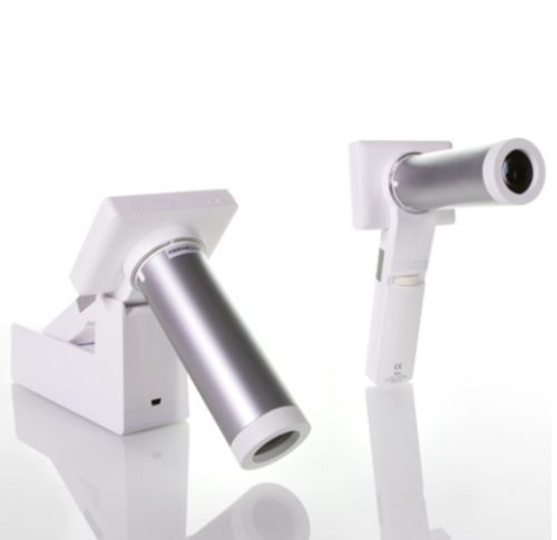
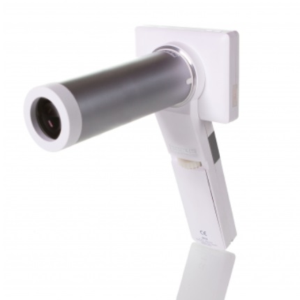
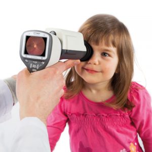
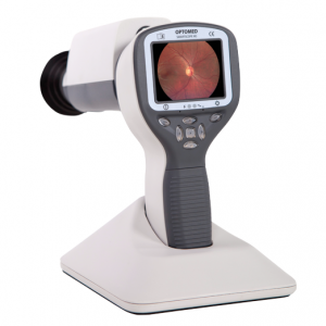
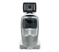