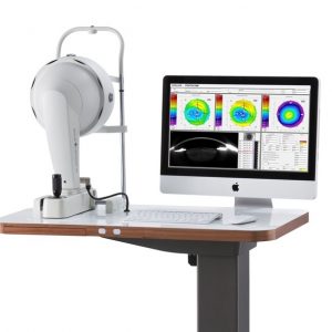
A Picture Says More Than 1,000 Words
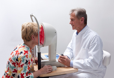
Use the OCULUS Keratograph® 4 as a marketing tool and incorporate it actively into your consultations. With the OCULUS Keratograph® 4 software, you can show images which your clients/patients have never seen before.
Contact Lens Fitting and Fluorescein Image Simulation
Contact lenses are recommended on an individual basis and displayed in a list. In order to avoid taking more steps than necessary when fitting contact lenses, the fluorescein image can be simulated beforehand. The contact lens can be rotated and moved around. Fluorescein image simulation is adjusted automatically. The integrated and expandable database contains all customary types of contact lenses and is updated on a regular basis. The user can determine the order in which contact lens manufacturers appear.
OxiMap® – Visualizing the Oxygen Transmissibility
Professional patient consultation
The cornea needs oxygen and good oxygen supply is fundamental for the comfort of a contact lens wearer. New materials used for soft contact lenses offer excellent oxygen transmissibility. This can be shown with the new OCULUS OxiMap® display. You can easily show these color maps to your patients and help them choose better contact lenses.
TF-Scan Makes the Tear Film Visible
This software shows the quality and quantity of the tear film.
In cases of dry eye patients and contact lens wearers, the tear film should be examined carefully. Only an intact tear film guarantees contact lens wearing comfort! The OCULUS Keratograph® 4 measures the tear film breakup time non-invasively (quality assessment). You can show your patient the individual tear film quality using the color maps and by taking another.
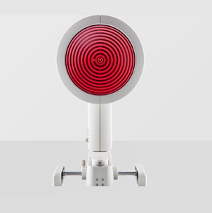
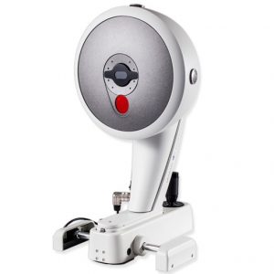
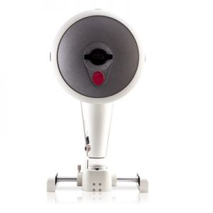
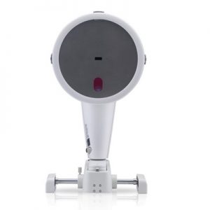
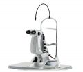
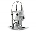
Technical Data
Features
| Measurement range |
3 – 38 mm, 9 – 99 D |
| Precision |
+/– 0.1 D |
| Reproducibility |
+/– 0.1 D |
| Number of rings |
22 |
| Working distance |
80 mm |
| Number of measuring points |
22,000 |
| Camera |
digital CCD camera |
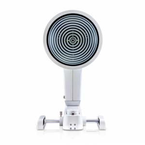
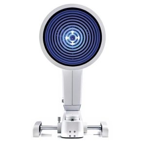
| Source of illumination |
placido illumination: red 650 nm
imaging illumination: blue 465 nm (UV-free)
pupillometer illumination: infrared 880 nm |
| Dimensions (H x W x D) |
49 – 51.7 x 27.5 x 32 – 40 cm |
| Weight |
15 lbs. |
| Minimum PC requirements |
Processor: Intel Core i3 or better, 4GB RAM, Hard drive space: min. 500 GB, Graphic card: Intel HD Graphics 2000 or better, recommended screen resolution: 1920 x 1080 (Full-HD) |

In acc. with the Medical Device Directive 93/42/EEC
 OCULUS is certified in acc. with DIN EN ISO 13485
Software
TF-Scan Makes the Tear Film Visible
Patient consultation made easy. This software shows the quality and quantity of the tear film.
In cases of dry eye patients and contact lens wearers, the tear film should be examined carefully. Only an intact tear film guarantees contact lens wearing comfort! The Keratograph® 4 measures the tear film breakup time non-invasively (quality assessment). You can show your patient the individual tear film quality using the color maps. In addition, you can take another non-invasive measurement to determine the amount of tear film (tear film quantity).
Tear Film Quality (NIKBUT)
The OCULUS Keratograph® 4 determines the break-up time using the NIKBUT procedure (non-invasive-Keratograph-break-up time).
Pupillometry
Using the “Pupillometry” option, the reaction of the pupil, can be checked with and without blinding. This builds the basis for selecting the proper treatment zone for laser controlled surface ablation, multifocal contact lenses or premium IOLs. The pupil reaction of the two eyes can be compared.
Optional Imaging-Software*
The Imaging-Software takes real-time fluo-images or fluo-clips of the contact lens. The high resolution clip shows the movements of the contact lens on the cornea. A side-by-side comparison displays the simulated fluo image of the software and the real time fluo image and the clip.
Essential for:
- Demonstration of the fit and mobility of contact lenses
- Assessment of the static fluo-image
- Assessment of the fit of the contact lenses with different pupil diameters
- Side-by-side comparison of fluo-image simulations to real-time fluo-images
- Choice of the final contact lense
- Expertise and customer long term relationship
OCULUS OxiMap®
The OxiMap® presents a color map of the oxygen transmissibility of soft contact lenses based on the lens power, which is easy to understand – even for your customers!
Documents
DICOM Conformance Statement for Keratograph®, 2.3.14
Download
DICOM Conformance Statement for Keratograph® 4, 1.76
Download